Inflammatory Disorders of the Esophagus: Reflux and Nonreflux Types


Inflammatory Disorders of the Esophagus: Reflux and Nonreflux Types


GASTROESOPHAGEAL REFLUX DISEASE (REFLUX ESOPHAGITIS-GERD/GORD)


Definition of GERD

Gastroesophageal reflux disease (GERD) is defined as at least weekly heartburn, acid regurgitation, or both, or extraesophageal symptoms with impairment of quality of life.1 GERD is quite common and affects around 20% to 30% of the population in industrialized countries. In the United States, 21% to 44% are said to suffer from regular heartburn, while GERD is actually diagnosed in 10% to 20%, with mucosal breaks (endoscopic erosions) being present in 2% to 5% of all refluxers. Around one-third of the affected individuals seek professional help. It is now clear that lifestyle factors, in particular obesity and smoking, are associated with increased reflux symptoms.2 In overweight individuals, the presence of a hiatal hernia increases the risk of reflux disease about ninefold compared to individuals without hiatal hernia.3

GERD is readily diagnosed in most patients from the characteristic history, and is treated by medications that decrease the amount of gastric acid. These range from over-the-counter antacids to medications reducing gastric acid secretion, of which there are two major classes. H2 receptor antagonists (H2RA) block the secretion of acid by blocking the histamine 2 receptors (hence H2) that mediate acid secretion in response to gastrin (released from the gastric antrum and duodenum) or acetylcholine. They usually have the suffix -tidine (cimetidine, ranitidine, etc.). Proton pump inhibitors (PPIs), as their name implies, inhibit secretion of hydrogen ions (protons) from parietal cells, and tend to be more potent although a little slower to act. They usually have the suffix -prazole (omeprazole, esomeprazole, lanzoprazole, etc.). Response to therapy is usually almost immediate, and further serves as a confirmation of the diagnosis.

Some patients have periodic symptoms and can take medications as the need arises, but for many it is chronic, so medications may be lifelong. Some patients become aware of the increased quality of life with PPIs when they undergo Helicobacter pylori eradication therapy, of which a PPI is a key ingredient. When the course of therapy ceases, their symptoms quickly recur so they become “hooked” on PPIs. While these are a remarkably safe group of drugs, they do increase the risk of gastrointestinal (GI) infections, and in the long term, gastric fundic gland polyps, which fortunately appear quite harmless. PPIs may increase the risk of osteopenia and fractures. In patients on longterm high-dose PPIs, the risk of vitamin B12 deficiency is increased, as both acid and B12 are required for its absorption, and the secretion of both from parietal cells is diminished by PPIs. Endoscopy, potentially with biopsies, is therefore reserved for patients who are not responding to therapy or have atypical symptoms such as chest pain in which it is unclear whether this is of cardiac, esophageal, or some other cause. While endoscopic erythema, erosions (mucosal breaks), and ulcers can all be seen, patients may have GERD without these changes (nonerosive reflux disease [NERD]), although on biopsy a variety of abnormalities can be found in these patients. Some of these patients appear to simply have exquisite esophageal sensitivity to acid.

There are numerous other causes of esophageal ulceration, especially medication, which may become stuck in the esophagus (pill-induced esophagitis), so that a history of medications used is also key (discussed subsequently). Infections (herpes, cytomegalovirus [CMV]) can also be painful and cause ulcers while there are numerous other less common causes (Table 10-1) that can affect the esophagus. Further, GERD has numerous complications but especially Barrett’s esophagus (BE), which increases the risk of esophageal adenocarcinoma, usually in association
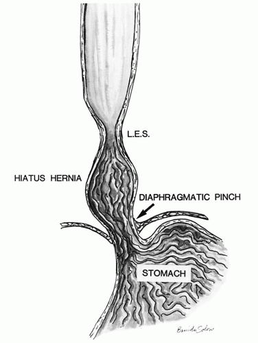
with hiatal hernia, which tends to promote free gastroesophageal reflux, as the additional safety valve of the diaphragmatic pinch is less effective. Other patients have motility problems that allow reflux of gastric contents into the esophagus or have a degree of peristaltic failure.


Table 10-1 Classification of Esophagitis by Etiology


1. Reflux—acid or alkaline reflux


2. Infectious


Fungal (e.g., moniliasis, Mucor)


Bacterial (e.g., mycobacteria)


Viral (e.g., herpes, CMV, HPV, HIV, varicella zoster)


3. Ingested substances


Medications


NSAID/ASA


Tetracyclines


Bisphosphonates (antiosteoporosis drugs)


Chemotherapy


Iron and Plummer-Vinson syndrome (ulcer due to iron-sulfur preparations for anemia)


Chemicals (acute)


Ingested acid or alkali


Silicates/metals/porcelain dust


Extremely hot or cold drinks


4. Trauma


Iatrogenic tubes (naso-gastric, pH-metry)


Bolus—injury due to foreign bodies


Radiotherapy


Ulceration of anastomosis (ischemic or insufficiency)


5. Systemic diseases


Collagenous diseases


Crohn’s disease


Sarcoidosis


Eosinophilic esophagitis


6. Tumors and their complications


Luminal obstruction


External compression


Necrosis


Neoangiogenesis


7. Miscellaneous


Exfoliative (sloughing) esophagitis


Symptoms of GERD

The leading symptom of GERD is heartburn, which needs to be differentiated from other causes of retrosternal pain, such as dysphagia, odynophagia, and cardiac pain. Further, GERD can lead to extraesophageal symptoms such as hoarseness, laryngitis, dyspnoea, cough, bronchitis and reflux-induced asthmatic disease, dental erosions, and retrosternal pain that need to be differentiated from ischemic heart disease.1 The frequency of these extraesophageal symptoms is probably underestimated in the population, but on the other hand laryngitis as a sequel of GERD is probably overestimated.4

Correlation between GERD, symptoms, and endoscopy. An ill-defined proportion of patients clearly have reflux-induced esophageal lesions endoscopically, but are asymptomatic, while another group have symptoms but little to see endoscopically (nonerosive [endoscopy-negative] reflux disease—NERD/ENRD).5, 6 A more troublesome group of patients appear to have persistent symptoms despite being on high-dose PPIs but have nothing evident endoscopically. These patients may have “visceral hypersensitivity” to esophageal acid, possibly mediated through dilated intercellular spaces (DISs) allowing acid (and other molecules) to pass through the pericellular space into the lamina propria and the nerves there.7 DISs are readily visible on biopsies, but it is unclear where physiology stops and pathology starts.

Therapy. In classical reflux disease, symptoms are treated with antisecretory therapy, usually PPIs.

A 1-week course of PPI that proves to be therapeutic can serve as a diagnostic test, and endoscopy is limited to those that do not respond, or have atypical symptoms, so that biopsies are not usually necessary.

Long-term therapy for GERD and prognosis. If longterm (usually lifetime) treatment with PPIs8 is refused, there are intraluminal endoscopic procedures available besides open or endoscopic fundoplication. What all of these procedures have in common is that they narrow the distal esophagus to imitate the distal esophageal sphincter to avoid regurgitation of gastric contents into the esophagus. Many of these endoscopic procedures have fallen out of favor because of either failure or complications. The surgical equivalent of these techniques has a small but definite mortality, related to some extent on the experience of the surgeon. However, patients who benefit most from a fundoplication are also those who show the best response to medical treatment. Also, fundoplication does not last forever (they become lax over time) and ultimately about 60% of operated patients need PPIs to manage symptoms. Approximately 20% of individuals who undergo surgical fundoplication may suffer from a gas-bloat syndrome where there is difficulty with eructation and air feels trapped and some develop dysphagia secondary to the fundoplication wrap being too tight, and dilation is required. General health (losing weight, sleeping with bed head raised) recommendations do not have long-lasting benefits in reflux disease. Nevertheless, there are always patients who report symptom relief after losing weight or after changes in lifestyle.


The Gastroesophageal Junction and Z-Line

One major problem is the difficulty in the definition of the gastroesophageal junction (GEJ), which differs worldwide. Practically by far, the most important definition is that used endoscopically in which the GEJ is now defined as the upper limit of the proximal gastric folds. Endoscopically, the Z-line is defined as the transition of columnar epithelium into stratified squamous epithelium. The Z-line is usually irregular (hence Z-line) and should be within the lower esophageal sphincter (LES), which itself extends for several centimeters (see Chapter 9). Histologically, squamous mucosa may be in direct continuity with:

1. Gastric cardiac, cardio-oxyntic, or less frequently oxyntic (acid-producing) mucosa

2. Undermining cardiac glands below the squamous epithelium (that can also be seen in treated GERD)

3. The presence of multilayered (hybrid) epithelium

4. Pancreatic metaplasia

The last three are usually not recognizable endoscopically.

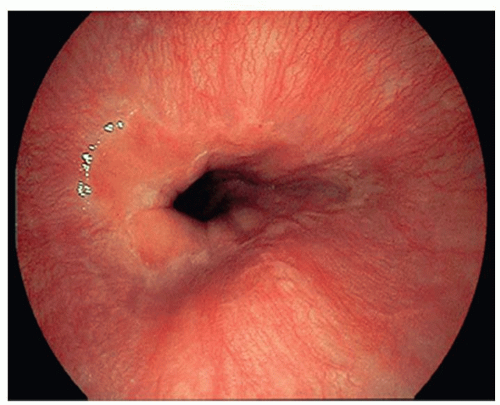
In Japan, the GEJ is defined by the distal end of esophageal palisade vessels. However, this is an approximation, as the end of these vessels can be present above, at, and even below the Z-line in normal individuals.


Etiology and Pathogenesis of GERD

Temporary mild reflux of gastric contents into the distal esophagus is thought to be physiologic. Pathologic reflux is diagnosed clinically whenever, in the supine position, a prolonged contact of gastric refluxate is present combined with a pH <4 for more than 4% of 24 hours (at least 1 hour a day). It is believed that low basal lower esophageal sphincter (LES) pressure
along with increased transient LES relaxations (tLESR) cause reflux disease. Besides increased relaxation, or insufficiency of the distal esophageal sphincter, there are other factors that can either cause or unmask prior existing reflux disease. These include the presence of a hiatal hernia (Fig. 10-1), the increased acid secretion that often follows H. pylori eradication therapy (or rarely Zollinger-Ellison syndrome), obesity, medications that decrease the LES pressure,9 possibly excess alcohol, impairments of esophageal emptying (motility disorders), impaired salivary and esophageal gland secretions, duodenogastric reflux, delayed gastric emptying (mostly in diabetic patients with autonomic neuropathy), stenosis or strictures, impaired esophageal resistance to acid, and failure of clearance. Numerous changes occur in the esophageal nerves (described under morphologic changes in nerves).

Gastric heterotopia (inlet patch) in the proximal esophagus can contribute to acid secretion, depending on its size and the presence of oxyntic mucosa.

For NERD, dilated intercellular spaces (DISs—see subsequent discussion) and visceral hypersensitivity are postulated mechanisms. The DIS experimentally can be induced by acid plus sodium nitrite,10 which is present in saliva.11, 12 In addition, the presence of duodenal contents containing bile and activated pancreatic juice, both known to be considered as etiologic factors in generating BE, might also be involved in the etiology of reflux disease. In large-scale studies, alcohol and smoking appeared not to be risk factors for reflux disease or its complications. General recommendations such as reducing weight may not impact reflux disease but in some contribute to reduced symptoms and of course make sense for general health.


Figure 10-1. Sketch of a sliding hiatus hernia. The LES is located in the chest above the diaphragm. (Courtesy of E. Hassall, MD.)

The role of hormonal effects is controversial: Gastrin and cholinomimetics increase the lower sphincter pressure, whereas secretin, glucagon, cholecystokinin, and prostaglandins inhibit the effect of gastrin.13


Endoscopic Grading of Reflux Disease in Squamous Mucosa

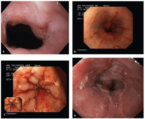
If endoscopy is carried out, there are numerous schemes available, but the Los Angeles classification (Fig. 10-2)14 is the best-validated classification scheme and is the most frequently used classification. It grades the extent of erosions and ulcers, which are together called “mucosal breaks.” A mucosal break is defined as a lesion covered by slough and is sharply delineated from the adjacent epithelium, often showing increased vascularity at the margin.

Grading is undertaken with letters A to D to avoid confusion with other classifications. Two additional grades were also proposed that would incorporate changes found in nonerosive reflux disease (NERD— discussed subsequently) with minimal changes (M) and without visible minimal changes (N)15 but these are not widely accepted or used outside of Japan. However, in common with the other classification systems, the Los Angeles system does not recommend taking biopsies.


Los Angeles Grading System for Reflux Disease in Squamous Mucosa

The Los Angeles classification (Fig. 10-2A-D) grades changes in squamous mucosa as follows:


Grade A—One or more mucosal breaks (= erosions) no longer than 5 mm, none of which extends between the tops of the mucosal folds

Grade B—One or more mucosal breaks more than 5 mm long, none of which extends between the tops of two mucosal folds

Grade C—Mucosal breaks that extend between the tops of two or more mucosal folds, but which involve <75% of the esophageal circumference

Grade D—Mucosal breaks that involve at least 75% of the esophageal circumference


Figure 10-2. Reflux esophagitis: Endoscopic appearance of Los Angeles A-D reflux esophagitis. (Courtesy of Steffen Muehldorfer, MD.) A: Grade A: A mucosal break <5 mm long between the tops of the mucosal folds at the right side of the esophageal wall. B: Grade B: Six mucosal breaks more than 5 mm long between the tops of the mucosal folds. C: Grade C: Mucosal breaks that extend between the tops of two or more mucosal folds, but involve <75% of the esophageal circumference. D: Grade D: Mucosal breaks that involve more than 75% of the esophageal circumference.

Proposed additions that would precede grades A to D15 are also occasionally used:


Grade N— Reflux disease with normal endoscopic appearance and absence of minimal signs and mucosal breaks

Grade M— Minimal changes other than mucosal breaks

In Europe, the Savary-Miller classification is often used.16 However, there have been several modifications, and interobserver variation is considered poor. The MUSE classification (Metaplasia, Ulcer, Stricture, Erosion)17 is very logical but not used.

For routine clinical practice, in endoscopic examination, the esophagus in patients with GERD, only five things are relevant:

1. Are erosions (called mucosal breaks in the LA system) present?

2. Is stenosis present?

3. Is BE present?

4. If BE is present, the detection of dysplasia/neoplasia.

5. Are subtle/minimal features present (redness, red streaks) that might reflect NERD?

Endoscopic grading is of less interest in a clinical routine setting, but erosions (mucosal breaks) may be relevant,
while stenosis may need to be dilated and BE might need PPIs (even if asymptomatic) and follow-up surveillance.


Nonerosive Reflux Disease and Its Pathology

It is now accepted that the vast majority of patients with reflux disease do not present with esophageal lesions (mucosal breaks/erosions) but do suffer from reflux symptoms.1 These patients are believed to belong to a group of patients with nonerosive (endoscopynegative or with minimal changes) reflux disease.6 However, using magnifying zoom endoscopy (magnification between 110 and 150 times), it has been shown that minimal lesions that do not fulfill the criteria of a mucosal break that occur in this group of patients are for the most part dilated intraepithelial capillary loops in the lower esophagus.18 Further, some patients may just be hypersensitive to esophageal acid.

This group can further be subdivided into true normals and those with minimal lesions such as vascular ectasia, an increase in DISs, and an increase in basal cell height or papillary height that is not in the pathologic range yet decreases considerably on PPIs. This suggests that NERD may consist of both patients with minimal lesions that can be demonstrated with appropriate endoscopes and a further subgroup of patients who may be very sensitive to acid (visceral hypersensitivity) but who are completely normal endoscopically.19 In both groups of patients, PPIs cause all of these changes to disappear, and the normal palisaded vessels at the Z-line become visible again, while many of the hypersensitive groups also become symptom free. The diagnosis of endoscopic-negative reflux disease is still somewhat difficult. pH-metry is considered to be the “gold standard” but is uncommonly carried out, although it can be done with a portable device at home.


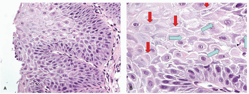
Figure 10-3. Dilated intercellular spaces. A: Overview of an esophageal biopsy with overt basal cell hyperplasia. DIS tends to be maximal around the papillae. B: Detail showing DIS that can take the form of exaggerated intercellular prickles (ladders) between any two cells (red arrows), or “bubbles” at the junctions of three cells (blue arrows).

Histology can help in this respect, depending mostly on the site of the biopsy. Biopsies close to the GEJ often show typical features of GERD but in biopsies close to the Z-line these probably have to be interpreted as normal, even though in some it represents genuine reflux disease as there may well be some differences between normal and abnormal (see previous discussion18, 40, 41).

The other features that can be found in patients with NERD are DISs (spongiosis) of the epithelium. This accentuates the intercellular prickles (“ladders”) between two adjacent cells, but at the intersection between three cells these form intercellular canals that appear rounded (bubbles) (Fig. 10-3). This intercellular edema is a good marker of epithelial damage, so it is not specific and is also common in eosinophilic esophagitis (EoE). In addition, small amounts of intercellular edema are common and probably normal, so when normal becomes pathologic is unclear. Care must be taken to not misinterpret cytoplasmic vacuolation as DIS.


Gross/Endoscopic Appearances of GERD

Endoscopically, early signs of reflux disease are for the most part found in the distal 2 cm of the esophagus (see Fig. 10-2), likely being most marked between the right and posterior wall of the esophagus.20 Within the valleys of the mucosal folds, esophageal gland ducts open and secrete bicarbonate and acid-neutralizing mucous. The top of the mucosal folds are therefore likely to be damaged mechanically during esophageal contractions with higher mechanical pressure, as well as by acid. If severe enough erosions form, in the Los Angeles system these are called “mucosal breaks.” Minimal changes that are not covered by this definition are not further defined in classifications on reflux esophagitis and are considered to represent their own group, rather than classified as true endoscopic-negative reflux disease.21 One example
is that of red streaks of the distal esophagus. These are covered by a thin layer of newly re-epithelialized squamous epithelium with underlying capillary-rich granulation tissue.21 Frequently, such lesions can be seen after healing of erosions (mucosal breaks).

Biopsies (see subsequent section) can be useful whenever atypical symptoms or questionable lesions are present endoscopically to exclude neoplasia. Changes may also be present in endoscopically normal mucosa that can support the clinical diagnosis of reflux disease.8


Where to Biopsy for GERD and Criteria Used

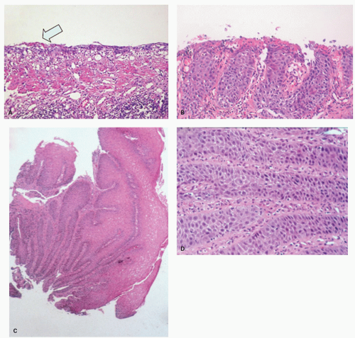
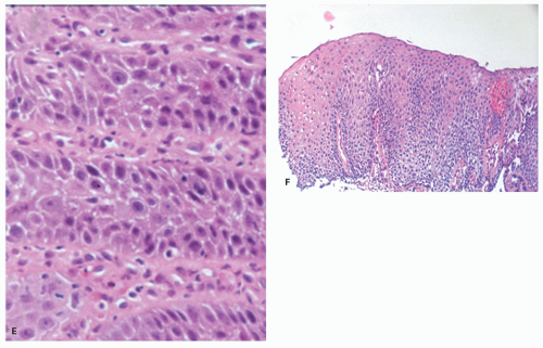
Erosions. Although erosions are not often biopsied to confirm their presence if they are multiple or atypical, consideration may be given to exclude other treatable causes such as Candida or herpes. Biopsies from erosions are easy to interpret when at the edge of a biopsy, as they consist of granulation tissue, sometimes with atypical stromal cells. However, small fragments may show only basal cells and underlying capillaries representing granulation tissue that is being re-epithelialized, some of which correspond to the red streaks seen endoscopically (Fig. 10-4A). Marked reactive changes may also be problematic as they may be confused with invasive squamous carcinoma. Initially they are part of re-epithelialization (Fig. 10-4B-E). The regularity of these “prongs” is the key to their reactive nature. More usually there are typical reactive changes with overt basal cell hyperplasia and papillary elongation (Fig. 10-4).


Figure 10-4. Erosions and sequela. A: Erosion with a wisp of superficial restituting epithelium (left arrow). Lesions such as these can be seen when red streaks seen endoscopically are biopsied. B: Erosion undergoing re-epithelialization. The epithelium consists entirely of basal cells dipping into the underlying granulation tissue. C-E: Healing erosion with marked pseudoepitheliomatous hyperplasia. The distinction from neoplasia at low power is the regularity of the “prongs.”

If no erosions are seen, biopsies from just above the Z-line can show evidence of healing erosion (Fig. 10-4),
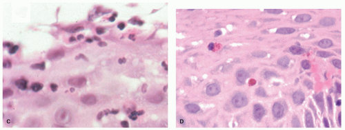
neutrophils that are initially superficial (Fig. 10-5), and a sprinkling of eosinophils (Fig. 10-5) that are less than those seen in EoE (<25 in 1 high-power field [HPF] or 15 in each of 2 in the same biopsy, and usually far fewer than this) but are usually sufficiently uncommon that one usually has to search for them. There is an overlap with EoE (see that section); patients with typical clinical, endoscopic, and histologic features of EoE are usually treated as such but where these are not all present (and in some patients with typical features of EoE), they are often treated for GERD, at least initially. A rare eosinophil is thought to be acceptable in adults, although they remain uncommon, but in children eosinophils are always regarded as abnormal. An excess of intraepithelial lymphocytes (IELs) (lymphocytic esophagitis) is seen in GERD but appears
to be neither sensitive nor specific; nor has the cut-off for normals been well defined. As such, this cannot reasonably be used to make a diagnosis of GERD.


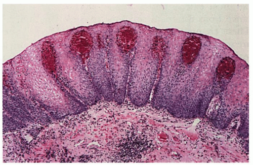
Figure 10-4. (Continued) F: More typical reactive changes in which the acanthotic epithelium has basal cell hyperplasia and papillae extend about 80% of the way to the surface. To the right there is a hemorrhagic papilla and the remnants of granulation tissue.


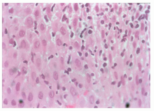
Figure 10-5. GERD. A: (detail C) Esophageal mucosa with superficial neutrophils. While this can be seen in GERD, it can also be part of drug-induced injury and infections such as Candida.


Figure 10-5. (Continued) B: (detail D) Epithelium with scattered eosinophils indicative of GERD.

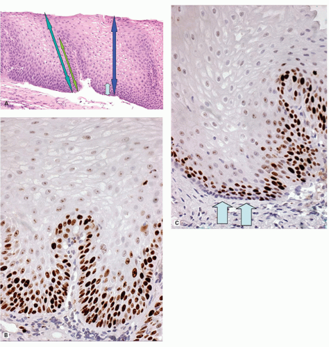
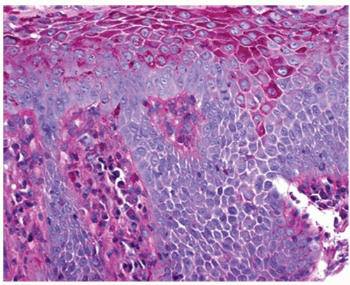
Biopsies 3 cm above the Z-line may show reactive epithelial changes (basal cell hyperplasia, elongation of papillary length) (Fig. 10-6). Biopsies also exclude imitations of reflux disease (Table 10-2) even if there are minor changes such as red streaks or areas of redness,22 and to recognize BE with or without dysplasia or carcinoma. Theoretically, a periodic acid-Schiff (PAS) stain can also be used to delineate the basal layer as these cells are glycogen deficient (Fig. 10-7), but this is rarely used as the hyperplasia is obvious on H&E sections (see Figs. 10-4F and 10-8). Another feature of basal cells is that they have a high proliferative index (see Fig. 10-6). In comparison, not only does normal mucosa have a much lower proliferative index, but in a Ki-67 immunostain the basal layer is unstained, but this disappears as soon as proliferation increases, suggesting that these may be part of the stem cell population of the esophagus.

Traditional biopsy sites. Distal esophageal biopsies are usually taken at the Z-line or 3, or 5, or 7 cm above the Z-line (depending on the author), but are said to play a minor role in diagnosis because in early papers basal cell hyperplasia and papillary elongation may be seen physiologically in the distal 3 cm.23 The closer these are taken to the Z-line the more likely these reactive changes are assumed to be present. It is likely that most of the regenerative changes are found between the right esophageal and the rear esophageal wall20 due to the asymmetrical structure of the LES. This seems to be the region where the valve-like mechanism of the LES opens first.24 However, this does not take into account that the other changes associated with GERD are all maximal close to the Z-line, so this is where biopsies should be taken to detect erosions or their healing phase (often red streaks), neutrophils, eosinophils, and markedly DIS even when the mucosa appears absolutely normal. Thus biopsies from this region should show more changes compared to other quadrants.

In the squamous mucosa, biopsies for features of GERD should therefore be taken

1. immediately above the Z-line for erosions, neutrophils, eosinophils, and DIS, preferably from the right esophageal wall (basal cell hyperplasia or papillary hyperplasia in this zone have to be interpreted as being within normal limits).

2. 3 cm above the Z-line for traditional reactive changes in the squamous mucosa.

3. from the mid or upper esophagus when EoE is in the diagnosis, hopefully to demonstrate their proximal persistence in that disease.

Cardia biopsies. A little-used criterion is the presence of inflammation limited to the cardia. The rationale for taking biopsies from the cardia is that there are only two major causes of cardiac inflammation: that associated with H. pylori and that associated with GERD. However to establish the latter, it is necessary to also take standard biopsies for Helicobacter from the gastric antrum and body—the latter are particularly important if patients are taking PPIs, and organisms may only be found in oxyntic mucosa. In the absence of inflammation in the remainder of the stomach, cardia inflammation is assumed to be secondary to GERD.25 A similar theoretical argument can be made with the incidental finding of intestinal metaplasia in cardiac biopsies; this should either be associated with intestinal metaplasia in the distal stomach, and therefore part of more widespread
gastric intestinal metaplasia, or limited to the cardia, and be GERD related.


Figure 10-6. Reactive changes in squamous mucosa. A: Normal squamous epithelium with small layer of basal cells extending about one-sixth of the distance to the surface (light blue and dark blue arrows right). The epithelial papillae do not extend more than about two-thirds of the entire epithelial thickness. This can be gauged by dividing the epithelium into thirds (light green and dark green arrows left). The papillae should not extend into the upper one-third, while if the basal third is mentally divided into thirds, the basal layer should not occupy more the half of this. The basal layer stops where the distance between nuclei is greater than the nuclear diameter. B,C: Ki-67 immunostain in reactive hyperplasia (B) and normal mucosa (C). In normal mucosa (right) the proliferative zone is above the most basal layer (arrows) suggesting that the nonproliferating cells are “stem cells.” In reactive changes (left) they are all reincorporated into the proliferative zone.


Histologic Diagnosis and Criteria for GERD in Biopsies

The following criteria are used in the morphologic diagnosis of reflux disease; however, as with inflammatory diseases elsewhere, no criteria are specific for reflux disease, and can be seen in other diseases also, causing erosions and ulcers such as infections (e.g., Candida, herpes simplex virus [HSV], cytomegalovirus [CMV]), medications causing ulceration (NSAIDs, bisphosphonates, etc.) that tends to be higher in the esophagus, although not often enough to be relied on, and rarely other causes (e.g., Crohn’s disease). (See subsequent discussion.):

1. Erosions or ulcers, usually close to the Z-line (other causes: infections, medication)

2. Re-epithelializing erosions (regenerative papillae elongation with attenuated overlying epithelium, and then with marked hyperplasia of the basal layer) (see Fig. 10-4)


Table 10-2. Reflux-independent Esophageal Lesions


NONNEOPLASTIC LESIONS WITHOUT REFLUX DISEASE


NEOPLASTIC LESIONS WITHOUT REFLUX DISEASE


Erosion


Drug-induced, mechanical (probes), ischemia, moniliasis, bacterial and viral infections, eosinophilic esophagitis


Early carcinoma


Systematic diseases with esophageal involvement


Dysplasia


3. Neutrophils (same differential diagnosis as 1) (see Fig. 10-5)

4. Eosinophils (differential is primarily EoE if numerous) (see Fig. 10-5)

5. Reactive epithelial changes—basal cell layer >15% to the surface or papillary height >two-thirds of the way to the surface (3 cm or more above the Z-line only)

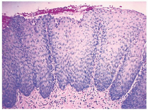
Evaluating reactive changes. In practice, no one measures reactive changes other than in research protocols, but the “rule of thirds” works well. The mucosa is divided mentally into thirds. Papillae extending into the upper one-third are abnormal. The basal one-third is mentally halved. Basal cells extending into the upper third of these are abnormal (in practice, 16.7% of the epithelial thickness, but close enough). The upper end of the basal cell layer is where the distance between the nuclei exceeds their diameter that is, where it becomes possible to fit in a nucleus between nuclei (see Fig. 10-5). This can also be demonstrated using the presence of glycogen (PAS stain) (see Fig. 10-7) that accumulates in cells above the basal layer, but this is rarely used. In a well-orientated biopsy, both basal cell hyperplasia and papillary elongation can be evaluated with relative ease (see Fig. 10-8).


Figure 10-7. PAS stain showing PAS-positive glycogen-containing cells that are not present in the basal cells and are limited to the nonbasal superficial cells.


Figure 10-8. The dermal papillae are elongated, and the basal cell zone is expanded to more than 15% of the squamous layer’s thickness. If these changes were found in a biopsy that included the Z-line, or were stated as being within 3 cm of the Z-line, it is appropriate to say that “reactive changes are present but in this location they may be physiologic.”

Less Reliable Criteria

1. Increased peripapillary hemorrhage (increased fragility of capillaries) (Fig. 10-9)

2. Presence of so-called balloon cells (acid-damaged squamous cells)

3. Eosinophilic “densification” of superficial squamous epithelium

4. DISs (likely a sensitive marker of any sort of epithelial damage but no good data on how much is normal)

5. Increase in IELs

Standardized criteria have been difficult to achieve as few of the criteria are specific for reflux disease, although very sensitive markers of esophageal damage.23
In practice, the most relevant (specific) criteria are thickness of the basal cell layer (>15%) and proportional length of papillae (>2/3),26, 27, 28 the latter invariably coming out as the best criteria in clinical studies. This may be useful as it suggests that the presence of an erosion or ulcer without immediately adjacent reactive changes in the squamous mucosa may be an indicator of acute damage (pills) rather than more chronic intermittent disease (reflux).


Figure 10-9. Dilated vessels in papillae (vascular “lakes”).

Dilated Intercellular Spaces It is clear that DIS (spongiosis) are a very sensitive marker of esophageal damage, and are seen especially in GERD, EoE, and “pill”-associated damage. However, the literature is relatively vague on criteria for their use. It has been suggested that although this initially referred to dilatation of the round spaces where pericellular spaces between more than two cells join (“bubbles”), this can also be applied to the spongiotic zone between two cells (“ladders”).29 Further, a degree of DIS can always be seen in the basal and prickle layers, so a small amount of this is always normal.

The question then is when can this be regarded as pathologic. In one study using oil immersion and electron microscopy, mean diameters were 0.58 ± 0.16 µm for controls, 1.07 ± 0.30 mm for NERD, and 1.29 ± 0.20 µm for erosive disease. The optimal cut-off value from receiver operator characteristic analysis was 0.85 µm.30, 31, 32 There are also data showing that DIS get smaller following antisecretory (proton pump) therapy, but this happens to some extent in controls as well as in patients, suggesting that DIS are largely acid reflux related.30 Anecdotally, we have found that numerous markers of intercellular junctions can facilitate demonstration of DIS including desmocollins, desmogleins, cadherins, catenins, occludins, and claudins. However β-catenin, which is available in many laboratories, works well and reliably until one can routinely identify these changes at routine H&E light microscopy.

Grading reactive epithelial changes. The original criteria for reactive esophageal squamous changes (basal cell hyperplasia, papillary elongation)26 were subsequently graded by Elster,33 which is difficult if biopsies are not well orientated, but grading has proven useful in routine practice. The system was


Grade I: The thickness of the basal cell layer reaches up to 10% of total epithelial thickness, papillae reach up to 40% of total epithelial thickness (i.e., normal).

Grade II: The basal cell layer reaches up to 30% (abnormal) and papillae reach up to 60% of total epithelial thickness (still just normal).

Grade III: The basal cell thickness is more than 50% and papillae reach up to 90% of total epithelial thickness.

In virtually all studies where both of these criteria have been measured, papillary elongation (height) is the most reliable criterion. A potential weakness of this system is that using papillary height, the abnormal range is in grade III, whereas using basal cell hyperplasia it is in grade II.

Signing Out Reactive Changes in Biopsies In routine practice, no criteria really need to be graded, although frequently that is the case, as it is almost second nature for pathologists to add “mild, moderate or marked/severe” as a prefix to anything that is a variable criterion. At the mild end, the pathologist may be trying to be helpful in providing a diagnosis. However, if the squamocolumnar junction is included in the biopsies then unless there is evidence of an erosion, all changes based entirely on the morphology of the squamous mucosa may be physiologic. While it is usually easy to say that basal cell hyperplasia or papillary elongation or marked dilatation of intercellular spaces (together often called “reactive changes”) are present, the issue is in their interpretation. It is not incorrect to say that they are “consistent with reflux disease,” as this may be true. However, this can be frankly misleading as they are also quite consistent with being within normal limits. One approach is simply to state that “reactive changes are present but these are normal in this location.” Of course, the additional presence of occasional neutrophils or eosinophils changes this immediately, but any change is based entirely on the presence of these cells, the changes in the squamous mucosa being irrelevant. Conversely, if it is clear that the biopsies come from 3 cm or more above the squamocolumnar changes, then they are pathologic, although a degree of caution is required because reactive changes can have numerous causes other than GERD, especially medications, as well as infection. Caution should therefore be exercised before mindlessly stating that reactive changes are “consistent with GERD” as it may be overtly misleading.

EROSIVE ESOPHAGITIS. In erosive esophagitis, the mucosa is reddened, the lesions are covered by debris, and whitish slough is found. Histologically, the margin of esophageal lesions shows the most marked regenerative changes reaching into hemorrhagic or necrosis with marked active inflammation including neutrophils, eosinophils, and less frequently lymphocytes and plasma cells. By definition, erosions do not extend into the submucosa while ulcers do. However, as the lamina propria underlies the epithelium, it is possible to completely lose the epithelium without satisfying the pathologic definition of an ulcer. This is usually impossible to assess in biopsies that may show only hyalinized lamina propria or granulation tissue
(Fig. 10-10), so it is usually assessed more precisely endoscopically.


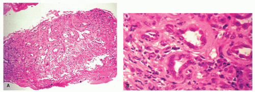
Figure 10-10. Exuberant granulation tissue in ulcerative esophagitis can mimic adenocarcinoma. A: Low-power view of a biopsy specimen from an esophageal ulcer. B: High-power view shows the microvessels of the granulation tissue.

Interestingly, although it would be reasonable to think that GERD-associated ulcers occur in association with reactive changes in the adjacent squamous mucosa, as opposed to no reactive changes, our own experience is that this is not the case, and that in biopsies from clinical studies from patients with erosive esophagitis who have a well-controlled drug history, that GERD-associated ulcers are frequently accompanied by a virtually normal adjacent mucosa. Further, biopsies from “mucosal” breaks may show a variety of changes from genuine erosions and ulcers through reactive changes to inflamed and normal mucosa. Whether the latter represents inadequate sectioning through the block or an endoscopic “miss” is unclear; the esophagus is constantly moving with respiration and heartbeat so is not the easiest organ to biopsy accurately. Therapy with PPIs is the therapy of choice.
INFLAMMATORY CELLS

Neutrophils are usually only found close to lesions, but can be helpful in biopsies from the Z-line when reflux disease is by far the most common cause. Neutrophils are usually found in the apical layer of squamous epithelium (see Fig. 10-5A-C), although when they start forming clusters it is wise to carry out special stains to exclude moniliasis, which can be easily missed on an H&E section.

However, asthmatics can also have reflux disease.34


Intraepithelial Mononuclear Cells

These are primarily lymphocytes (CD3+, usually CD8+) with a varying portion of S-100-positive antigen-presenting cells, but mast cells can also be present. Intraepithelial mononuclear cells (IEMs) are eosinophils that can be observed at the margin of esophageal lesions (especially pill-associated ulcers with homogenous eosinophilic necrosis) but, apart from a rare cell, are not found in normal squamous epithelium. Scattered eosinophils (see Fig. 10-5B-D) imply GERD until proven otherwise. In children, they are always assumed to be abnormal. In this respect, EoE should always be excluded. Asthmatics can have an increased number of intraepithelial eosinophils, possibly related to the asthma therapy increased in reflux disease (Fig. 10-11), but there are no controlled studies available that help in grading this finding.35 Although there is an association with GERD and numerous other disorders including inflammatory bowel disease, it is wise to be cautious about interpreting an esophageal lymphocytosis (vide infra section on lymphocytic esophagitis).


Figure 10-11. Esophageal mucosa with myriad IEMs, most of which are lymphocytes, but antigen-presenting cells and sometimes mast cells may all be present.


Minor changes

Nerves Numerous neuropeptides and changes in them are reported in reflux disease (such as CGRP, neuropeptide Y, Substance P, and VIP). Generic neural markers (such as PGP 9.5) have been evaluated in the esophageal epithelium. For VIP and PGP 9.5 a relation to inflammation and thus symptoms in reflux disease has been shown.36 The relevance of these findings is not yet known. Increased capsaicin receptor TRPV1 (transient receptor potential cation channel, subfamily V, member 1) nerve fibers have been found in the inflamed human esophagus, but the potential diagnostic utility of this finding is unclear.37 In the mouse esophagus, capsaicin inhibits the vagally mediated striated muscle contractions mainly through its action on mucosal primary afferents, which in turn activate the presumed inhibitory local reflex arc.38 Inhibition of contractions would promote reflux. Cholinergic and nonadrenergic noncholinergic inhibitory nerves that are nitric oxide mediated play important roles in regulating contraction and relaxation of the human LES. A decrease of the action of cholinergic nerves and an increase of the action of nonadrenergic noncholinergic inhibitory nerves by nitric oxide may be largely related to the low LES pressure observed in patients with reflux esophagitis,39 while increased serum levels of both nitrous oxide and VIP that mediate esophageal inhibitory neurons are elevated resulting in relaxation.40 Nitrous oxide is also implicated in the DISs associated with GERD.12 It may cause relaxation of the LES and, in the presence of acid, result in DIS that are also the direct result of reflux.10, 12, 41 Further, DIS may also be potentiated by direct central vagal stimulation.42


Inflammatory Polyps

These are by far the most common polyps in the vicinity of the Z-line, and follow erosions and ulcers irrespective of the etiology. Most begin as granulation tissue but are then re-epithelialized by mucosa that may come from squamous mucosa, or are the glandular mucosal types found at the Z-line. However, squamous mucosa is by far the most common. There are several potential diagnostic pitfalls with these polyps:

1. Pseudoepitheliomatous hyperplasia.

2. The cuboidal-like endothelium can appear almost epithelial, so that their random nature may be mistaken for invasive carcinoma. This is particularly likely if frozen sections are carried out (see Fig. 10-10).

3. Atypical reactive stromal cells (pseudosarcomatous changes in the stroma) (Fig. 10-12). These have a range of appearances but can look overtly malignant, but are always single, stain only with vimentin, sometimes CG10, while Ki-67 can vary from entirely negative to most cells staining, although when the latter occurs, mitotic figures, which can be atypical, are invariably present. Because these cells are seen in inflammatory polyps, invariably adjacent to erosions or ulcers in a variety of locations, they are sometimes colloquially referred to as “ulcerocytes,” which at least gives them the harmless name they deserve. The trap is to regard these as spindle cell carcinomas, or “sarcoma” in an inflammatory polyp.


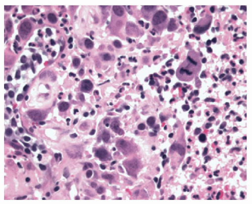
Figure 10-12. Reactive (pseudosarcomatous) changes in the stroma. Note the lack of cohesion, large nuclei but with indistinct chromatin, and, in this example, mitotic figures. These cells are immunoreactive to vimentin, sometimes CD10, and occasionally Ki-67.

Reactive changes versus neoplasia following erosions and ulcers. There are two main circumstances where this is a problem:

A. Distinguishing basal cell hyperplasia from squamous dysplasia

B. Distinguishing the marked hyperplasia of rete pegs from invasive squamous carcinoma

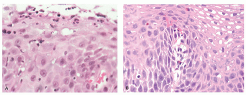
Basal Cell Hyperplasia versus Squamous Dysplasia A side-by-side comparison with low-grade dysplasia is shown in Figure 10-13. with low-grade dysplasia Typically, squamous dysplasia ranges from mild to in situ carcinoma (low- to high-grade dysplasia [HGD]/intraepithelial neoplasia [IEN]). The nuclei are usually open, vesicular, contain more chromatin, are punctate, and small nucleoli may be present. It is usually fairly regular throughout the biopsy although in resections junctions and different grades may be present. There is invariably loss of polarity in the basal layer so the nuclear features of one nucleus cannot be readily predicted from its neighbor. Whenever this issue arises, it is always worth ensuring that there is no another cause for the epithelial hyperplasia, especially a granular cell tumor or fungal infection, so both are worth a search.


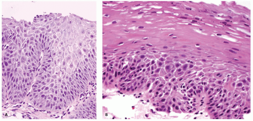
Figure 10-13. Reactive hyperplasia (A) versus low-grade squamous dysplasia (B). Note the orderly maturation in the squamous mucosa but disorderly maturation, sometimes with almost an abrupt transition from immature to mature epithelium as shown here. A helpful clue is that other features of reactive changes (papillary hyperplasia, extensive dilatation of intercellular spaces) are present on the left, but rarely present in squamous mucosa. Although dilated intercellular spaces are present in the dysplastic mucosa on the right, they stop abruptly where the squamous dysplasia stops.

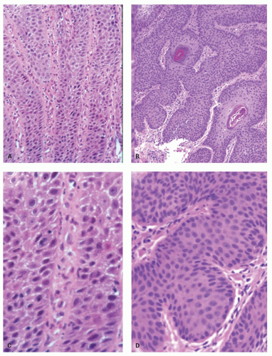
Marked Hyperplasia of Rete Pegs versus Invasive Squamous Carcinoma In this the regularity of the rete pegs which is orderly is contrasted with the disorder and randomness of the neoplastic pegs that is the key to this diagnosis. This is also best illustrated (Fig. 10-14).

Perhaps the most difficult of these is when verrucous carcinoma is in the differential diagnosis. These are also discussed in Chapter 11, but dysplasia/atypia in the basal layer may be the only feature that allows this distinction morphologically short of invasion, which can be quite subtle. It also differs from reactive changes in that the architecture is also not predictable. Rete pegs in carcinomas are irregular, cannot be predicted, can have little or no orientation to the point that one assumes the biopsy is very poorly oriented, and when well oriented, the pegs vary in size and shape with some often being club shaped amidst others that are tapering or anastomsing. The second feature is that the nuclei are different in that they are more open, vesicular, may have small nucleoli, and, at least in the basal layer, there is a lack of predictability with loss of polarity. However, the latter may be limited only to the basal layer, and even within it may be focal, although readily identified. Although usually HPV related, HPV effects may be present but may also be focal; similarly in situ hybridization for HPV may also be focal. If p16 is strong and diffuse that should help, but it is usually weak or absent, even in those associated with HPV, usually subtypes 6 or 11. Sometimes one gets the impression that reactive changes are admixed with verrucous changes. The saving grace in this situation is that the biopsy invariably comes not from a possible ulcer or erosion but from an endoscopic lesion. The presence of an endoscopic tumor should also raise the possibility that one is dealing with an inflammatory polyp, especially close to the GEJ. These are relatively common. Verrucous carcinomas are also discussed in the following chapter.


Atypical Cells in Squamous Mucosa

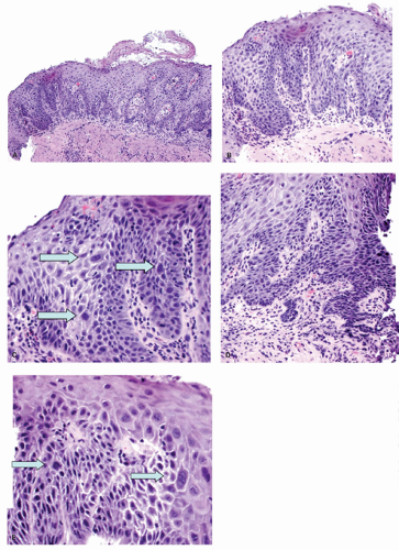
A degree of individual nuclear changes can be seen in changes that are not overtly reactive or dysplastic, but these are better demonstrated than described (Fig. 10-15).


Carditis as a Manifestation of GERD

Several studies suggest changes even distal to the Z-line can be part of reflux disease. Apart from H. pylori, there does appear to be an association between GERD and inflammation limited to the gastric cardia.25, 43 In these studies, active and chronic inflammation was only found in patients with reflux disease (neutrophils, lymphocytes, and plasma cells). Unfortunately, these findings could not be reproduced in larger studies.44, 25 It is not clear yet whether cardia mucosa is a metaplastic mucosa per se.45 Conversely, fetuses and children do have cardia mucosa.46, 47


Figure 10-14. Reactive versus neoplasia. The panels on the left (A,C) show orderly reactive hyperplasia where the squamous rete pegs (prongs) are orderly and predictable in their regularity. In comparison, in this example of one of the best-differentiated squamous carcinomas we could find (B,D), the major difference is at low power (A,B) where the tumor grows in a disorderly and unpredictable manner. The details (C,D) show dysplastic mucosa to primarily have greater cellularity. Loss of polarity, especially in the basal layer, together with lack of reproducibility between adjacent cells, and greater hyperchromatism in dysplastic mucosa tend to be the best features.

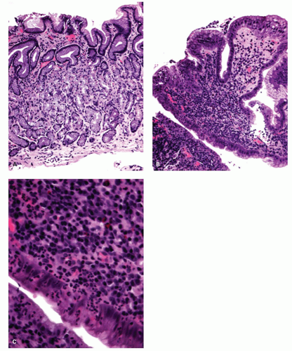
For routine diagnosis, the presence or absence of carditis seems not to be in standard use, possibly because it is necessary to not only take biopsies of the cardia, but also do the usual two antral and two oxyntic mucosa biopsies to exclude Helicobacter as a cause, for if these are present, the presence of carditis cannot be used to confirm the diagnosis of reflux disease, as the carditis is presumably Helicobacter related. Is it possible to have carditis in the absence of both GERD and Helicobacter? To date only one study that we are aware of has suggested that this might occur.44 The combination of normal (Helicobacter-negative) biopsies from the gastric antrum and corpus, along with chronic or chronic active cardia inflammation, needs an explanation. In the absence of other explanations, it seems very likely that these are GERD related (Fig. 10-16).


Figure 10-15. Atypia in squamous mucosa. A: Overview of lower esophageal biopsy showing marked nuclear changes, especially in the papillae that extend very close to the surface. Note that the epithelium rests almost on top of the muscularis mucosae with virtually no lamina propria, suggesting that this is a re-epithelialized erosion. B,C: Details of the left end of (A), with enlarged atypical nuclei (arrows). D,E: Adjacent field from (A) showing the restituting edge of an erosion lower right (E), immediately adjacent mucosa to (D), with further individual atypical cells, the largest (right) in E being binucleate (arrows). Situations such as these are best handled by treating with PPIs and repeating the biopsies within a month or two. Endoscopy should be carried out with high a resolution endoscope, biopsies being directed at atypical areas.


Figure 10-16. Chronic active carditis. A: Antral biopsy with no hint of Helicobacter gastritis (key in determining that this is the case, as antral and cardiac mucosa tend to resemble each other in Helicobacter infection). B: Biopsy from cardia— same patient and same magnification as “A” showing marked inflammation. C: Detail of (B) showing the chronic active inflammation. In the differential diagnosis, the severe chronic inflammation makes medication use unlikely and Crohn’s disease is unlikely as a presenting manifestation. This essentially leaves GERD or an “idiopathic” etiology. The former seems the most likely choice. Diagnosis was “chronic active carditis that, in the absence of Helicobacter, is most likely GERD-related.”

Progression of GERD. In adults, there appears to be almost no progression of reflux disease with time, although in children the situation is unclear. Over time, repeated healing and damage of the epithelium can be noted but progression from endoscopically negative to erosive and to BE seems not to occur—or if it does it is appears to be uncommon.48 The more accepted model of reflux disease is therefore a steadystate model. Furthermore, it is unknown why there is much variation between patients, with some patients showing alterations of the very few distal centimeters and others showing lesions 15 cm and more above the GEJ.


Complications of GERD

These include ulcers that can penetrate but do not perforate the esophageal wall. Fistulas can occur between esophagus, bronchus, pericardium, or aorta but are extremely rare, although they do occur in squamous cell carcinoma. Strictures are for the most part a sequel of long-standing ulcerative reflux disease, but are becoming rare as a result of sufficient acid suppression. Severe hemorrhage is also rare but bleeding can lead to chronic iron deficiency.

Reflux-induced neoplasms are always adenocarcinomas associated with BE. The risk for the
development of carcinoma is 40- to 125-fold higher in BE compared to in the normal population (see following Section and Chapter).

GERD-related strictures. Strictures are usually managed by endoscopic dilatation, but occasionally need to be resected. They invariably occur immediately proximal to the Z-line and are 1 to 4 cm long. When resected, the stricture itself may be ulcerated, and the stricture is seen to be due to marked submucosal fibrosis and smooth muscle proliferation, the latter penetrating extensively into the muscularis propria. Immediately proximal to the stricture the muscle is markedly hypertrophied, and the epithelium may be thickened and hyperplastic. Reactive changes are present at the junction of ulcerated and nonulcerated mucosa; residual esophageal glands are often visible in the submucosa, and sometimes in the lamina propria.


Cameron’s Ulcer

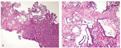
This is an ulcer, usually longitudinal, but can involve much of the circumference of the mucosa, that occurs in a hiatal hernia as it goes through the diaphragm, and so is found at that constriction. These ulcers can bleed, and be associated with hematemesis or anemia. The etiology of these ulcers is not known, but the few that we have seen have been surprisingly severe, sometimes almost purulent but without identifiable organisms, and sometimes with an overt ischemic component (Fig. 10-17).

There are many postulated mechanisms, but if these ulcers by definition occur at the diaphragmatic hiatus, this is likely involved in its pathogenesis. If there is an ischemic element, then some form of pressure-induced ischemia seems most likely. In patients taking appropriate medications, pill damage needs to be considered, as these can cause ischemic-type damage and injury might be caused if they are held up at the diaphragmatic pinch. Other factors that have been implicated inevitably include gastric acid exposure, although this is likely to potentiate or prevent healing in established ulcers rather than being the primary cause. PPIs therefore tend to be used in their therapy and appear effective, presumably confirming a role for acid, unless this is a placebo effect. Gastric stasis and lymphatic obstruction have also been implicated.


Figure 10-17. Cameron’s ulcer in a hiatal hernia. A: Oxyntic mucosa (right) with an exudate on the surface (left). B: Detail of part of the pseudomembrane showing pits with attenuated restituting epithelium in a fibrin neutrophil exudate and bearing considerable resemblance to pseudomembranous colitis. Although no fibrin thrombi are present in this biopsy, its apparent acute ischemic nature is apparent.

A potential clue to the diagnosis in biopsies is that because these are in a hiatal hernia, accompanying squamous mucosa is absent in the ulcerated fragments, the background mucosa being cardiac or oxyntic. However, it needs either knowledge of the clinical location or recognition of a potential ischemic component in the biopsy, or both.


BARRETT’S ESOPHAGUS (BE)


Practical Aspects of Making the Diagnosis of BE

In many countries, including much of North America and Europe, clinicians and pathologists are much more confident in making the diagnosis of BE if goblet cells are present. The presence or absence of goblet cells in biopsies should therefore be stated, if only for traditional reasons.

Nevertheless, the definition of BE still requires two components,

1. demonstration of a proximal shift in the Z-line endoscopically, and

2. histologic demonstration that the mucosa that has shifted proximal is columnar (with or without goblet cells depending on the country).

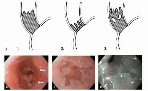
Usually the presence of a proximal shift in the Z-line is apparent endoscopically as tongues, circumferential spread, and occasionally squamous islands as judged by proximal extension from the endoscopic landmarks (proximal limit of the gastric folds, although
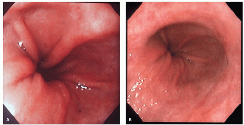
recognition of the diaphragmatic pinch can also be useful, especially in establishing the presence of a hiatal hernia that invariably accompanies BE (Fig. 10-18), but when the squamous mucosa is inflamed it becomes red, so the proximal limit of the Z-line may not be apparent, and it is here that biopsies may be required to confirm that the proximal mucosa is indeed columnar and not squamous. The top of the gastric folds marks the proximal limit of the stomach (Fig. 10-19), although this is not as easy as it seems, as it depends in part on the degree of insufflation.


Figure 10-18. Endoscopic appearances of BE. A: Sketch illustrating the patterns. A common pattern is shown in panel 1. The interface between the Barrett’s (hatched) and squamous mucosa is often wavy, as shown, similar to the Z-line at the GEJ. Panel 2: Short segment with tongues only, that is, no circumferential involvement. Panel 3: Residual whitish squamous islands are commonly present, especially near the upper end of Barrett’s mucosa. B: Endoscopic view of long-segment Barrett’s with circular columnar metaplasia and small islands of squamous epithelium (arrows). C: Tongues of columnar metaplasia in the distal esophagus (BE) in white light. D: NBI technique with regular gyriform surface epithelium pattern. (Images courtesy of Steffen Muehldorfer, MD.)

The exception to this two-part practical definition is Japan, where the CLE is defined on endoscopy alone, by the distal limit of the lower esophageal palisade vessels53 (Fig. 10-20). However, use of these anatomic landmarks to define the length of BE results in a large number of patients with this condition, hence all patients with BE <5 mm in length, without any other factors associated with typical BE, are excluded.54, 55 It is unclear how this correlates with BE as defined above, and what proportion of these patients have intestinalization in this mucosa, and if so how much of this is otherwise typical cardia-type mucosa.

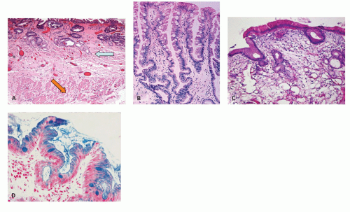
In much of Europe, intestinal metaplasia (i.e., goblet cells) is required on biopsy from the suspected segment of endoscopic BE to establish the diagnosis56 (Fig. 10-21A,B). In the United Kingdom, the presence of columnar metaplasia alone with or without goblet cells (Fig. 10-21C,D) in the distal esophagus defines BE, the assumption being that every columnar mucosa has goblet cells somewhere.51 The diagnosis of BE is therefore primarily endoscopic, biopsy being used to confirm the diagnosis and to ensure that, as this is also the first surveillance endoscopy, as far as possible dysplasia or carcinoma is not already present.

Irregular Z-line versus “ultrashort” BE. Apart from in Japan, the top of the gastric folds marks the beginning of the cardia, from which the Z-line has to be shifted proximally to make a diagnosis of BE. The most difficult endoscopic decision is where an irregular Z-line becomes a tongue of BE. In an interobserver endoscopic study of “experts,” there was no agreement until the tongue was 1 cm or more in length,54 reflecting the difficulties in reliably measuring columnar tongues <1 cm. The implication of
this is that it is impossible to make a confident diagnosis of BE until the proximal extension of columnar mucosa, whether a tongue or circumferential extension, is at least 1 cm in length. Less than this it is simply an irregular Z-line by definition. The corollary of this is that it is impossible to separate “ultra-short” BE (<1 cm of BE) from intestinal metaplasia in native cardiac mucosa. Although ultrashort BE presumably must exist, at the time of writing there is no good way to make that diagnosis by biopsy of this region alone. If intestinal metaplasia is isolated to the cardia region without the rest of the stomach being involved, this implies that it may well be GERD related and therefore Barrett’s related, but this remains implied and not definitive. The latter requires biopsies of that area most likely to have intestinal metaplasia, namely, the antrum and incisura region of the stomach, the most likely cause of which is Helicobacter, and which is mirrored at the cardia. If biopsies are carried out to ascertain this, those from the cardia must necessarily be placed into a separate container to allow identification of cardiac and antral pathology separately.


Figure 10-19. Endoscopic Landmarks: Recognizing the GEJ. A: Top of the gastric folds. B: LES pinch that is frequently lax in patients with GERD-associated disorders. Here it is at the top of the gastric folds.


Figure 10-20. Endoscopic landmarks of the distal end of the esophagus (in Japan) showing the limits of the palisaded vessels. In this example, they focally descend beyond the upper limit of the gastric folds.

With this proviso, intestinal metaplasia limited to the cardia (no metaplasia in the antrum or rest of the stomach) is likely to be reflux related.

Definitions of BE: short- versus long-segment BE. Endoscopically, BE has been divided into short segment (<3 cm) or long segment (3 cm+). This is a historical feature that is slowly losing favor due to lack of relevance. It was developed because it was known that the Z-line should be somewhere within the LES, which is about 3 cm long. Anything more than 3 cm was therefore “unequivocal” BE, whereas anything less than this was open to question. However, it was clear that some of the excluded patients also had to have BE, so the issue became how best to make that diagnosis, particularly when biopsies of that region showed that some of these patients also had intestinal metaplasia, the presumed precursor of esophageal carcinoma.


Figure 10-21. A: Histology of columnar metaplasia with goblet cells/specialized intestinal metaplasia and double muscularis mucosae. The marked and diffuse intestinalization in the lower esophagus is virtually diagnostic of BE as such metaplasia is only seen in gastric mucosa with diffuse intestinal metaplasia. In addition, the duplicated muscularis mucosae is clearly visible with a superficial (neo) layer immediately beneath the epithelium (blue arrow) and the original muscularis mucosae beneath (orange arrow). Courtesy of Dr. C Streutker. B: Typical biopsy appearances of BE with incomplete intestinal metaplasia and branching pits. C: Histology of columnar metaplasia without goblet cells. This patient had a 3.5-cm circular segment of columnar metaplasia in the distal esophagus with no significant hiatal hernia. Repeated endoscopy and multiple biopsies still did not show goblet cells. D: Alcian blue stain at pH 2.5. While the goblet cells stain, many of the intervening columnar cells also stain. Not only can interpretation be difficult but it can sometimes be positively misleading. We rarely carry out this stain for diagnostic purposes.


When BE Does Not Have Goblet Cells, Does It Matter?

The requirement of goblet cells for the diagnosis of BE is based on the notion that carcinoma only develops if intestinal metaplasia is present. Yet there are abundant data to suggest that the non-goblet cell population in BE already have most of the abnormalities found in goblet cells, including many of the characteristics of an intestinal phenotype,52, 57 and the necessity of having goblet cell required for the diagnosis of BE has been questioned.54

When are goblet cells not present? There are numerous circumstances when BE is present endoscopically but goblet cells are not found. These include the following:

1. Children with BE do not usually develop goblet cells until into the second decade of life.

2. Some patients with tongues of columnar mucosa do not have goblet cells when first biopsied. While some have them on repeat biopsy (presumably sampling problems first time), some never seem to develop them; do these patients have “a tongue of BE,” and if not, what does one call this appearance if not a tongue of BE? Perhaps just “a tongue of columnarlined esophagus.”

3. Several large studies appear to show that some patients with unequivocal BE endoscopically never seem to develop intestinal metaplasia. From our own unpublished data about 80% of segments <1 cm do not have goblet cells. With increasing length the probability of detecting goblet cells increases but never reaches 100% even in very long segments. It is not clear how much this reflects reality and what proportion is due to sampling error.

The first paper to suggest this was by Kim et al.,58 in a Veterans Administration study that included only patients with long-segment BE (>3 cm). They found that 20% of patients with BE did not exhibit intestinal metaplasia in biopsies from two endoscopies, a figure similar to that obtained by Harrison et al.59 over a decade later. Also, in the latter study, of 125 patients with BE, only 35% of patients would have been diagnosed with BE if only one to four biopsies were obtained; 68% if five to eight biopsies were obtained; and 74% if more than eight biopsies were obtained.

In another multicenter study looking at 3,568 nondysplastic biopsies from 1,751 patients, intestinal metaplasia was detected more commonly in males (odds ratio 1.24) and in patients with a longer columnar segment length.60 In that study, the chance of detecting goblet cells was also proportional to the number of biopsies obtained. About 55% of patients without intestinal
metaplasia at index endoscopy showed intestinal metaplasia after 5 years, while at 10 years the figure reached 91%. Thus, 9% of patients in these cohorts never had goblet cells detected over that time period.

Another quirk is that goblet cells may be easier to find in short- rather than long-segment BE as goblet cells are preferentially found proximally in columnar-lined mucosa. In one study, 70% of patients with 1 to 2 cm and 90% of patients with 3 to 4 cm of columnar metaplasia had goblet cells—more than that seen in many patients with long-segment BE. In this study, 94% of proximal biopsies had goblet cells compared with only 39% of biopsies from the distal esophagus.45 Obtaining biopsies from the distal esophagus only could have resulted in BE being missed in more than 50% of patients. There are therefore many reasons why goblet cells may not be detected: They may be present but not detected; they may not be present; they may come and go.

Risk of carcinoma in patients with non-goblet cell BE. It is difficult to decide what type of follow-up the individuals without goblet cells within columnar metaplasia should receive, but this should likely be the same as for those with goblet cells. The important thing is that if goblet cells are not found, that the patient is not lost to follow-up assuming they still have a risk of cancer. But do they?

Several studies have shown a similar risk of neoplastic progression in patients with BE, irrespective of the presence of goblet cells. Gatenby et al.60 looked at 322 patients with and 612 patients without intestinal metaplasia, and found that 19.8% of the former and 15.2% of the latter developed LGD or HGD or adenocarcinoma on follow-up (no significant difference).

Similarly, in another follow-up study by Kelty et al.,61 the rate of development of adenocarcinoma was 4.5% in those with intestinal metaplasia and 3.6% in those without. Putting both studies together, perhaps there is a slight additional risk if goblet cells are present, but it is also clear that the dictum “no goblet cells, no cancer risk” is incorrect.52, 62

Only gold members can continue reading. Log In or Register to continue

Stay updated, free articles. Join our Telegram channel

Jul 20, 2016 | Posted by in GASTROENTEROLOGY | Comments Off on Inflammatory Disorders of the Esophagus: Reflux and Nonreflux Types

Full access? Get Clinical Tree

Get Clinical Tree app for offline access