Immunodeficiency Disorders


Immunodeficiency Disorders


INTESTINAL HOST DEFENCES

The gastrointestinal (GI) tract must digest and selectively absorb nutrients, while at the same time excluding large amounts of potentially harmful ingested substances such as microorganisms and toxins.1, 2, 3, 4 Both immune and nonimmune defense mechanisms are important for intestinal host defense.3, 5, 6, 7, 8 This defense is provided within the lumen by secretory IgA (originating from the intestine or bile) and within the mucosa by lymphocytes (mucosal alpha-beta and gammadelta T cells),5, 6, 8 plasma cells, and macrophages.9 Secretory IgA has been likened to antiseptic paint, which lines the bowel mucosa, acting as a protective layer. It has four antigen-combining sites and is very efficient at agglutinating bacteria and viruses and preventing their adherence to mucosal surfaces. In addition, IgA interferes with the absorption of many macromolecules by combining with food. This helps prevent harmful systemic immune responses. The antigens (particulate and soluble products) that do escape the action of secretory IgA and penetrate the surface epithelium may form immune complexes, which can be cleared by the liver and excreted in the bile. Alternatively, they may be cleared by locally sensitized lymphocytes, by combination with preformed antibodies, or by ingestion by macrophages.5

Much of the work on gut immunology has centered on the role of humoral immunity on gut defense mechanisms. However, B-cell function is often under the control of T cells.5, 10, 11 Intraepithelial lymphocytes (IELs), most of which are of T-cell (CD-8) type, have an important cytotoxic action. Cell-mediated immunity appears to play an important role in
fungal infections such as candidiasis, certain viral diseases, and parasitic infections, whereas humoral immunity seems more important in protecting against run-of-the-mill enteric bacterial and viral infections.12

A variety of nonimmunologic factors contribute to gut host defense. They include the physical integrity of the mucosa/mucosal barrier (intestinal permeability); intestinal mucus, which may impair antigen binding and allow antigen degradation by intestinal enzymes; resident microbial flora; acid and pepsin, which cause bacterial and dietary antigen degradation; bile acids, which suppress microbial proliferation; and bowel motility. The latter produces regular cleansing of the intestinal tract.13, 14, 15


FUNCTIONAL ANATOMY OF THE GI IMMUNE SYSTEM


Normal Distribution of Gut-associated Lymphoid Tissue

Lymphoid tissue is normally abundant throughout the GI mucosa (including IELs) with the exception of the stomach and in fact is the largest lymphoid organ in the body.8 In the stomach, there are almost no lymphocytes and plasma cells within the gastric fundus and body and only a few within the antrum and cardia. The lymphoid tissue first appears in the lamina propria of the bowel at 10 weeks16 and by 14 weeks lymphoid follicles develop. Plasma cells appear at birth but are very scanty.16 The lymphoid tissue is arranged in three forms:


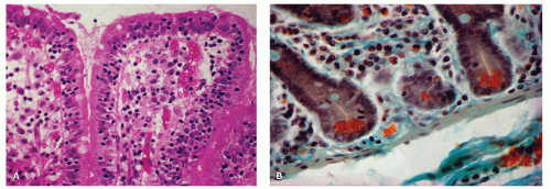
Figure 3-1. The diffuse lymphoid system of the intestinal tract. A: Intraepithelial lymphocytes (IELs). Mucosal biopsy specimen from a patient with tropical sprue, showing intestinal villi with an increased number of lymphocytes within the surface epithelium. B: Section of normal jejunal mucosa showing lymphocytes, plasma cells and eosinophils within the lamina propria between the intestinal crypts. Using this stain, Paneth cells in the crypt bases are bright orange red. (Masson’s trichrome stain)

1. As IELs, throughout the GI tract, including the esophagus.

2. As a diffuse lymphoplasmacytic infiltrate, which is distributed evenly throughout the intestinal mucosa of the small and large intestines.

3. As lymphoid nodules. There are essentially two types:

a. Solitary lymphoid nodules present throughout the GI tract but most numerous in the distal colon.17

b. Aggregates of lymphoid nodules, which occur in the appendix and small intestine. In the small intestine, these aggregates are referred to as Peyer’s patches and are most frequent in the distal ileum.18

The lymphoid tissue in the ileocecal valve is unique in that it is arranged circumferentially around the valve. The mesenteric lymph nodes are also usually considered part of the gut-associated lymphoid tissue (GALT). In common with the gut, mesenteric lymph nodes are exposed to considerable antigenic material via the lymphatic flow from the small and large intestines and are populated by predominantly IgA precursor B-cell lymphocytes.

The Diffuse Lymphoid Tissue This tissue is contained in two separate compartments, namely, intraepithelial and intramucosal.19

INTRAEPITHELIAL LYMPHOCYTES. These cells are located within the surface epithelial layer of the mucosa, the so-called IELs (Fig. 3-1A). They occur predominantly in the basal portion of the epithelial layer, between the epithelial cells and the basement membrane.20, 21
Although they appear to be few in number in any one section (at the range of 10-25 per 100 epithelial cells in the small bowel and ˜5 per 100 epithelial cells in the colon),22, 23, 24 when the entire intestine is considered, they are in fact very numerous and are said to equal in aggregate the number of lymphocytes found in the spleen. Histologically, the IELs consist of dense nuclei with minimal cytoplasm and do not have any epithelial attachment. The predominant phenotype is the cytotoxic T cell expressing αβ T-cell receptors, which are CD3+, CD8+, CD103+, CD4−, and CD5−. A smaller population (10%-15%) consists of T cells expressing γδ T-cell receptors that are negative for both CD4 and CD8. A third population of CD56+ IELs is also recognized that are virtually undetectable in normal mucosa. They have T-cytotoxic and natural killer (NK) properties.20, 25 The IELs overlying the lymphoid follicles consist predominantly of B cells. In addition, other intraepithelial cells such as macrophages, mast cells, neutrophils, and eosinophils may also be present.16, 23, 26 IELs are greatly increased in several diseases, such as celiac sprue, tropical sprue, lymphocytic colitis, and collagenous colitis. They are typically not increased in inflammatory bowel disease.23 In inflammatory states, neutrophils and eosinophils may also enter this compartment.27

Lymphocytes are also present within the esophageal squamous epithelium. They occur primarily within the suprabasal portion of the mucosa, interdigitating between the squamous cells, and phenotypically consist of CD3/CD8 cytotoxic suppressor cells.28

INTRAMUCOSAL CELLULAR INFILTRATE (LYMPHOCYTES, PLASMA CELLS, EOSINOPHILS, MAST CELLS, MACROPHAGES, ETC.). The cells consist primarily of numerous lymphocytes, plasma cells, and eosinophils. In addition, a heterogeneous group of other cells are also present in smaller numbers, namely, eosinophils, mast cells, macrophages, dendritic cells, rare basophils, and T lymphocytes (Fig. 3-1B). The latter consist primarily of helper inducer cells.29

Intramucosal lamina propria. With regard to lymphocytes, the lamina propria is the largest compartment of GI lymphocytes, the number being in the range of several thousands per square millimeter.

They are located primarily in the crypt region and less frequently in the villi in the small bowel. The majority of plasma cells, many of which contain Russell bodies, consist primarily of IgA-containing cells, followed by IgM, IgG, and IgE.25 IgD-containing plasma cells are exceedingly sparse in the normal GI tract.

Neutrophils are exceedingly rare/absent in the normal lamina propria. However, eosinophils are generally present, ranging up to 200 per mm2.30 The number of eosinophils in the colon is highest on the right side and lowest in the rectum. The number of eosinophils in the lamina propria also varies geographically according to latitude, as the farther north one goes in the United States, the fewer eosinophils one finds in normal colon biopsies.31

Mucosal mast cells. The mast cells occur predominantly in the lamina propria as well as the submucosa and appear to be more common in the ileum than the jejunum, up to about 750 cells per mm2.32 In the mucosa, they are primarily within the superficial half of the lamina propria. They have proved to be more numerous than was previously thought, mainly because they were previously missed in routinely prepared tissue sections. In animal studies, there is great heterogeneity among mast cells in that intestinal mast cells are morphologically and functionally distinct from peritoneal and systemic mast cells.33 The same type of difference may also exist in man, but this remains to be proven.34 Mast cells have physiologic regulatory effects as well as pathologic effects.35 Their function was previously thought to be confined to IgE-mediated hypersensitivity reactions. However, recent animal studies have shown that they are also involved in the late-phase components of allergic reactions, delayed-onset hypersensitivity, and regulation of immune responses. With regard to the functional activity of the mast cells, a variety of substances are released from the cells, leading to a number of factors such as blood flow regulation, endothelial and epithelial permeability, angiogenesis, mucosal secretion, and intestinal motility.32 Moreover, mast cells can express direct cytotoxic activity and can potentiate eosinophil and macrophage cytotoxicity.34 In certain conditions, such as nematode infections, gastritis, celiac sprue, idiopathic inflammatory bowel disease, and irritable bowel disease, mast cell numbers may be markedly increased in concert with other inflammatory cells.36

Histologically, Carnoy’s fixative37 was found to be better than formalin for identifying mucosal mast cells. Recently, however, a number of immunostains have been helpful in identifying these cells such as mast cell tryptase, CD117 (C kit),38 and CD68 (KP-1).39 These stains seem to obviate the need for any special fixatives as they work well on formalin-fixed tissue.

Mucosal lamina propria macrophages Macrophages are present in small numbers within the lamina propria, mainly beneath the top of intestinal villi and within the surface of the large intestine. Mucosal macrophages at the base of the lamina propria are generally less numerous, occurring primarily in the stomach and small intestine. Morphologically, they may be readily missed if not deliberately searched for. However, they increase greatly in number in inflammatory conditions. Functionally, they seem to play an important role in the intestinal mucosal immune system and also in inflammatory responses.40, 41


The Solitary and Aggregate Lymphoid Follicles (Peyer’s Patch)

SOLITARY LYMPHOID FOLLICLES. These are abundant throughout the small and large bowel but are most numerous in the distal colon.17 It has been estimated that there are several thousand follicles in the small intestine and up to 20,000 in the colon. The number of follicles within the stomach is very few. The structure and function of the lymphoid follicles appear to be similar to those of the Peyer’s patches.17, 42

PEYER’S PATCHES. Peyer’s patches are aggregated nodules of lymphoid follicles, which are most numerous in the distal ileum,18, 43 are located on the antimes-enteric border of the intestine, and measure from a few millimeters to 10 cm (Figs. 3-2 and 3-3).18, 44 The number of Peyer’s patches is variable and increases with age until puberty. There are approximately 240 at puberty.18 Thereafter, the number falls to about 100 by age 70 years.18


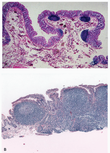
Figure 3-2. Solitary and aggregate lymphoid follicles of the human intestinal tract. A: Colonic mucosa and submucosa containing three solitary lymphoid follicles. The latter extend beneath the muscularis mucosae, and there is dimpling of the surface epithelium over the follicles. B: Peyer’s patch, composed of numerous lymphoid follicles aggregated together in a linear fashion.


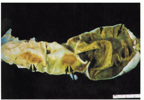
Figure 3-3. Autopsy specimen of terminal ileum and cecum. Two Peyer’s patches are visible in the ileum, measuring more than 3 cm in length (double arrows). Solitary lymphoid follicles are discernible in the cecum as tiny brown spots (arrow).

Morphologically, Peyer’s patches and lymphoid follicles have a distinctive structure. They are situated mainly in the mucosa but often split the muscularis mucosae and extend into the superficial submucosa. Three zones characterize them: (1) a dome region, (2) a follicular area, and (3) a parafollicular zone (Fig. 3-4).


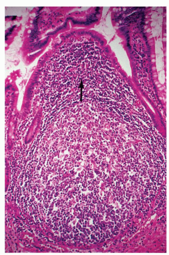
Figure 3-4. Intestinal lymphoid follicle. This is composed of a large follicular zone and the dome region, which is the area lying between the follicle and the surface epithelium (arrow). The mucosal surface overlying the follicle is dome shaped, devoid of villi and crypts, and lined by cuboidal epithelium.


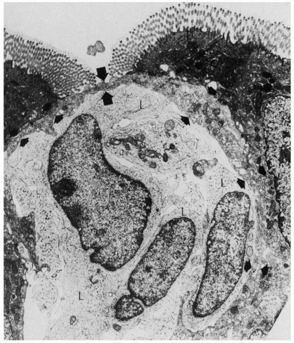
Figure 3-5. Electron micrograph of an M cell. This is a thin, membrane-like cell (arrows), the luminal surface of which is covered by microfolds and not microvilli. In the space below the M cell are three lymphocytes (L). (Courtesy R. L. Owen, M.D.)

The Dome Region This region lies immediately above the follicular area and is lined by a mucosal epithelium, sitting on a very porous basement membrane. This region essentially separates the lymphoid follicles from the lumen. The mucosal surface is dome shaped, relatively free of villi and crypts, and lined by a single cell layer. These cells are composed predominantly of cuboidal rather than columnar epithelial cells, a few goblet cells, the M cells (membranous or microfold epithelial cells), and occasional tuft cells.45 The epithelial dome cells essentially consist of specialized enterocytes migrating up from a separate crypt stem cell line.46, 47 The M cells predominantly lie between the columnar cells overlying the Peyer’s patches (and isolated follicles).48, 49 They are not readily visualized by light microscopy because of their size and are characterized ultrastructurally as thin membrane-like cells with luminal surface microfolds rather than microvilli (Fig. 3-5). These attenuated M cells form a latticework that allows lymphoid cells in the lamina propria to approach within 0.3 µm of the intestinal lumen while maintaining the integrity of the intestinal epithelium.50 M cells contain multiple vesicles that suggest a transport function of luminal antigenic material to the underlying lymphoid tissue.45, 47 Lastly, it should be noted that numerous groups of IELs are also present, composed of B cells and suppressor T cells. Furthermore, lymphocytes and mononuclear cells can easily migrate through the surface epithelium and basement membrane and frequently do so. It is important to recognize this as a normal histologic finding (this is not where one should assess the numbers of IELs if one is worried about celiac disease or microscopic colitis).

The subepithelial area contains many lymphocytes, plasma cells, macrophages, and dendritic cells. The lymphocytes are large and medium sized, consisting primarily of IgM+B cells and CD4 T cells and far fewer CD8 T cells. The macrophages not infrequently contain cellular debris and bacterial remnants. The dendritic cells, which appear to be bone marrow-derived cells of myeloid origin,51, 52 form a reticular network in the lamina propria. Functionally, the dendritic cells are potent intestinal antigen-acquiring cells with long dendritic processes that present antigens to T cells for induction of a primary T-cell response.52, 53 It has also been shown that the dendritic cells transport apoptotic intestinal epithelial cells to T-cell areas.53 In addition, some of the dendritic cells also appear to transport antigens to lymph nodes.52


Tissue subjacent to the dome region contains mainly small lymphocytes and scant macrophages.

Follicular Area This lies beneath the dome region and is composed of a follicle center surrounded by the mantle and then the marginal zone. The follicular center is similar to that within lymphoid follicles and is composed predominantly of dark-staining centroblasts at its base and mainly light-staining centrocytes in the center and mucosal surface. In addition, numerous tingible body macrophages, often containing debris, are present at the center of the follicle with scattered T-cell lymphocytes.42 The mantle zone surrounds the follicular center most prominently at the mucosal side and is composed of small B lymphocytes. Lastly, the outer portion of the follicle consists of a broad marginal zone composed of small- to intermediate-sized B lymphocytes containing a moderate amount of cytoplasm and centrocytic-type nuclei.42

The Parafollicular Zone This represents the thymus-dependent area and is characterized by small lymphocytes and many postcapillary venules, similar to those seen in lymph nodes. Under normal conditions, most of these lymphocytes are CD4 T-cell lymphocytes. However, in diseases such as inflammatory bowel disease, there is an increase in the number of B-cell lymphocytes with surface IgG.27

It should also be noted that prominent arterioles are present within the follicle and extend into the subepithelial capillary network and then to the capillary venules within the parafollicular T-cell zones. As far as lymphatics are concerned, they are prominent within the interfollicular and perifollicular areas and deal with lymphocytic migration.54, 55 Ultimately, lymphocytes migrate through the Peyer’s patch and flow toward the submucosal lymphatics. From there, they exit the intestine via the afferent lymphatics into the mesenteric lymphatic vessels to mesenteric lymph nodes and ultimately to the thoracic duct and peripheral circulation.

Immunohistochemistry of Peyer’s patches As far as the follicular centers are concerned, immunohistochemically they are similar to lymph node follicles and stain positively for all immunoglobulins other than IgD (primarily on the cell surface and less significantly in the cytoplasm). The follicular center cells consist of mature B cells that express CD10 antigen.42 The surrounding mantle zone B cells stain for IgM and IgD, whereas the marginal zone B cells are IgD negative but IgM and IgA1 positive, similar to splenic marginal zone cells. The parafollicular zone is predominantly composed of T cells, CD4 being about four times more common than CD8.

Humoral immune system of the gut. In man, five major immunoglobulins are normally synthesized and secreted by plasma cells, namely, IgG, IgA, IgM, IgD, and IgE.56 The differences in these immunoglobulins are due to the structure of their heavy chains. In tissue, there is a close correlation between the number of specific plasma cells and the synthesis of the corresponding immunoglobulins, and specific plasma cells usually predominate in different tissues. Thus, after the neonatal period, IgA-containing plasma cells predominate in the GI tract, and the corresponding immunoglobulin is found in the intestinal secretions.56, 57 The other immunoglobulin-containing cells are also present, albeit in lesser numbers.58 In the small intestine, it has been estimated that the ratio of IgA- to IgM- to IgG- to IgE-containing plasma cells is roughly 20:3:1:1.56 The estimates for the large intestine and appendix are marginally different, 21:1:5:1:1.59 IgD-containing plasma cells are exceedingly sparse in the normal GI tract.

Secretory IgA is the major immunoglobulin in the GI secretions, and most is produced by the intestinal IgA-producing plasma cells.57 Interestingly, animal studies have shown that a significant proportion of the secretory IgA found in the intestinal juices originates from the liver.60 The secretory IgA is elaborated within the biliary epithelium in a manner similar to that of the intestine,60 although it is not yet clear whether most of the IgA originates from the intestinal plasma cells via the enterohepatic circulation or partially from plasma cells resident in the liver. It has been estimated that about 3 g is produced daily, with a half-life of 4 to 7 days.61 Secretory IgA has a unique structure, which confers upon it the property of resisting degradation by intestinal enzymes. It is composed of two monomeric 7S IgA units held together by a protein, the J (joining) chain.62 In addition, IgA also contains a nonimmunoglobulin polypeptide chain, known as the secretory component.63 The dimeric IgA and the J piece are synthesized by the plasma cells of the lamina propria and are secreted into the interstitial space. They are then taken up by the intestinal epithelial cells, through the lateral cell membranes, where they acquire the secretory piece.63 The latter may be a structural component of the lateral epithelial cell membrane. Finally, the immunoglobulin is secreted by the epithelial cell into the gut lumen, where it forms a close association with mucins and coats the apical surface of enterocytes. The secretory piece is specific for IgA and gives it a unique property, namely, protection from proteolytic degradation by the enzymes of the gut, such as trypsin and chymotrypsin.64 There are two subclasses of IgA: IgA1 and IgA2. In the serum, IgA1 accounts for approximately 90% of the IgA, whereas in secretions IgA2 accounts for 60% of the total IgA. Other immunoglobulins are also found in small intestinal secretions and interestingly, IgM but not IgG follows a pathway similar to that of IgA.63, 65


Immunologic Events Leading to IgA Secretion One exciting development in gut immunology is the evolving concept of the mucosa-associated immune system (MALT), of which the gut-associated lymphoid tissue (GALT) is an important component (Fig. 3-6).7 This system confers local immunologic protection and, more importantly, is independent of the systemic immune system. The mucosa-associated immune system is present at all epithelial surfaces in contact with the external environment and is controlled by direct antigenic contact not only of the GI mucosa but also of other extraintestinal epithelial sites.10 This system is unique in a number of ways; the antibodies produced by the plasma cells are transported to the epithelial surface and are able to function in the presence of proteolytic enzymes. Furthermore, the locally activated lymphocytes have the unique property of homing back to their site of origin and other epithelia, such as lung, breast, genitourinary tract, and female genital tract, after maturation. Thus, local antigenic stimulation at one site, such as the GI tract, elicits an immune response not only in the intestinal mucosa but also at other mucosal sites, such as the urinary tract (Fig. 3-6). The immunologic process leading to IgA secretion involves intestinal and extraintestinal events.10 In animal studies, the first step involves sampling of antigens from the gut lumen by the M cells.64 Macromolecules are actively transported from the lumen through the cell to lymphocytes and macrophages lying in the tissue spaces immediately beneath these thin membranes. The antigens are then delivered to the Peyer’s patches where clonal proliferation of IgA precursor lymphocytes occur,64 probably under the control of helper T lymphocytes.11 IgA precursor lymphocytes next migrate to the mesenteric lymph nodes, thoracic duct, and systemic circulation, where they undergo maturation (possibly in the spleen) and then home back to the intestine and other mucosal sites,64, 65 where they populate the lamina propria, predominantly as IgA-synthesizing plasma cells (Fig. 3-6). Whether a similar mechanism is operative in man is uncertain.58 The activated lymphocytes of the bowel may lodge in other secretory organs, such as salivary glands, bronchial mucosa, and the mammary glands.64 The mammary glands are particularly important in the neonatal period in that passive transfer of IgA occurs via breast milk to confer protection to the newborn. It should be noted that the plasma cells of the GI tract develop only in response to exposure of foreign antigen within the intestinal luminal contents. Consequently, the fetal gut is devoid of plasma cells, the latter developing only after birth, following neonatal exposure to food.


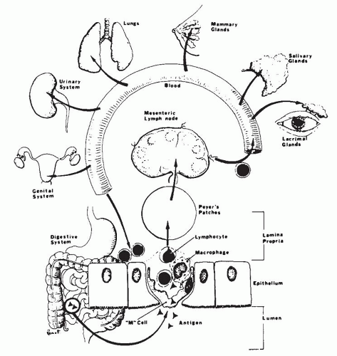
Figure 3-6. Mucosa-associated lymphoid system.

Cellular immune system of the gut. The lymphocytes of the cellular immune system (T-cell system) are an important component of the gut-associated lymphoid system. They are the major component of the IELs,6, 23, 26 and in the lamina propria they are distributed primarily in the interfollicular zone. The T-cell subtypes have a rather distinctive distribution, the suppressor T cells being located mainly in the epithelial compartment, while helper T cells are found in the lymphocytes of the interfollicular zone. The cellular immune system of the gut appears to be involved in the full complement of cellular immune functions, namely, recognition, processing, and destruction of antigens. Paneth cells contain a variety of substances that incude lysozyme, phospholipase, and defensins and also contribute toward innate immunity in the gut.


IMMUNODEFICIENCY DISORDERS OF THE INTESTINAL TRACT

The GI tract is frequently an important target organ in the primary as well as secondary immunodeficiency disorders66, 67 because it is constantly exposed to a heavy antigen load. Immunodeficiency is usually suspected by the clinician when patients have a history of either prolonged infections or infections caused by unusual organisms. The pathologist’s role is largely to confirm and identify the frequent complications of immunodeficiency, of which infection and neoplasia are the most important. On occasion, the pathologist may be able to document the type of immunodeficiency through the
use of immunohistochemistry. Sometimes, unexpected histologic changes may be found in the gut, such as nodular lymphoid hyperplasia and giardiasis, which should lead one to suspect an immunodeficiency disorder in a clinically unsuspected case. As common variable immunodeficiency (CVID) is so common, it may sometimes be detected in relatively asymptomatic patients because of a paucity of IgA plasma cells in the lamina propria with no compensatory increase in other classes of plasma cells.

There are two major categories of immunodeficiency disorders: primary and secondary.

Primary Immunodeficiency Disorders These disorders include a large and varied group of diseases66, 67 resulting from impairment of the B- and T-cell systems, abnormalities of phagocytic function, or both. The functional interaction of B and T cells and monocytes in the expression of the immune system makes the division of immunodeficiency diseases into B- and T-cell disorders somewhat artificial.6, 68, 69 Nevertheless, usually either the antibody defects or cell-mediated abnormalities predominate, allowing for a practical subdivision of the immunodeficiency disorders. In addition, genetic defects have been described in conditions such as food intolerance, in which there appear to be specific abnormalities in responsiveness to certain antigens. In these disorders, there appears to be poor antigen clearance or heightened responsiveness, leading to immune complex disease or hypersensitivity reaction.70 In adults, common variable or late-onset immunodeficiency, and selective IgA deficiency are by far the most common immunodeficiency disorders.15, 68, 71 In children, severe combined immunodeficiency disease is the most common. It usually presents soon after birth. Its diagnosis is important to prevent the sequelae of acute and chronic infection.2

Secondary Immunodeficiency Disorders These disorders are becoming more common. Some develop following infections, while others are iatrogenic complications of immunosuppressive therapy, radiation therapy, or bone marrow/hemopoetic stem cell transplantation (BMT/HSCT). The most important secondary immunodeficiency is acquired immunodeficiency syndrome (AIDS), which is caused by the human immunodeficiency virus (HIV).72 There is considerable evidence to suggest that immune mechanisms are involved in the tissue injury of a number of GI disorders, such as idiopathic inflammatory bowel disease, celiac sprue, and Whipple’s disease. The precise pathogenic mechanisms of these disorders remain to be elucidated and they are discussed further under each disorder.

Clinical features. Clinically, patients usually present with chronic diarrhea and malabsorption, although other symptoms may also occur based on the specific disorder (primary or secondary immunodeficiency) and its complications. The main impairment of the immune system is characterized by a decreased resistance to infection and in some cases, subsequent neoplasia.72, 73, 74 The nature of the infection depends on the specific defect. Thus, patients with impaired B-cell function are most susceptible to bacterial and certain parasitic infections. In contrast, those with impaired T-cell function have defective cell-mediated immunity, leading to sprue-like disorders, pernicious anemia, nodular lymphoid hyperplasia, inflammatory bowel disease,74 and an enhanced susceptibility to prolonged viral, fungal, and mycobacterial infections. The major clinical manifestations of these infections will depend on the primary site of involvement in the GI tract.75

The diagnosis of the primary immunodeficiency disorders is usually made readily. Patients commonly have a history of recurrent, prolonged opportunistic infections, such as pneumocystis pneumonia, giardiasis, or cryptosporidiosis. The finding of associated disorders, such as respiratory tract infections and autoimmune disorders in CVID, helps greatly in determining the precise immunologic abnormality and there is frequently a family history of immunodeficiency disease.

The categorization of the immunodeficiency disorders is based primarily on three categories of immunologic tests for humoral (B-cell) and cell-mediated (T-cell) immunity, that is, immunoglobulin concentrations, antibody formations, and cell-mediated immunity (Table 3-1).68, 69

Histology. The pathologic changes accompanying the primary immunodeficiency disorders depend in part on the specific immunologic defect (Table 3-2). It should be stressed that in some cases there may be no significant histologic alteration. The most frequently seen histologic lesions are the results of complications, specifically infections. Intrinsic lesions of immunodeficiency are uncommon. The histologic changes fall
into four major categories as described in the paragraphs that follow.


Table 3-1 Immunologic Tests for the Categorization of Primary Immunodeficiency Disease


  1. Immunoglobulin concentrations in serum and mucosal secretions.


  2. Assessment of antibody formation following immunization


    1. Natural antibodies such as A and B isohemagglutinins, and antistreptolysin


    2. Antibody response to immunization such as diphtheria/tetanus vaccine


    3. Quantitation of circulation B cells.


  3. Cell-mediated immunity.


    1. Skin testing for delayed cutaneous hypersensitivity with antigens such as mumps, trichophyton, PPD, Candida, and tetanus toxoid.


    2. T-cell quantitation including helper/suppressor ratios.


Table 3-2 Pathology of the GI Tract in Primary Immunodeficiency Disorders


No change


Specific morphologic changes


Altered lymphoplasmacytic content of lamina propria


  1. Absence of lymphocytes and plasma cells


  2. Altered B- or T-cell subsets


Diffuse nodular lymphoid hyperplasia


Nonspecific morphologic changes due to bacterial overgrowth, infections, or other injury


Esophagitis


Mucosal erosions


Atrophic gastritis


Mucosal lesions (villous atrophy)


Enterocolitis


Demonstration of organisms particularly


Candida, CMV, herpes, Giardia, Trichomonas, Isospora belli, Cryptosporidium


Neoplasia


Carcinoma


Lymphoma


Kaposi’s sarcoma


No Change There may be no significant alteration or reduced number of inflammatory cells of the lamina propria (Fig. 3-7). For example, in late-onset immunodeficiency disease, there may be an absolute marked reduction in plasma cells. Occasionally, plasma cells are not reduced markedly. Rather, there is a reduction of IgA-containing plasma cells with a compensatory increase in the number of the other plasma cells, especially those containing IgM. Immunostains will show the specific cell type that is altered, for example, a diminution in number or absence of IgA-containing plasma cells.


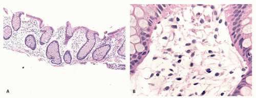
Figure 3-7. A: Large bowel mucosa with architectural distortion but an empty lamina propria. B: Detail showing almost entirely mesenchymal cells. Changes such as these can be seen in neonates and young children before the intestinal immune system has developed, in patients with immunologic disorders such as combined immunodeficiency disease, and also immediately following bone marrow transplantation before repopulation of the lamina propria has occurred. It can also be seen in some patients with inflammatory bowel disease following therapy and in remission.

Mucosal Lesions Resulting from Infections or Bacterial Overgrowth Infections in patients with immunodeficiency disorders tend to be more prolonged and cause more damage in the immunocompromised setting. Patients are especially prone to infections by multiple organisms, frequently of low virulence, which are not normally pathogenic to man. These include the normal commensals of the GI tract,76 fungi such as Candida,77 and viruses such as Cytomegalovirus (CMV) and herpes.68, 71, 76, 77 The patients are also unable to mount an effective host response to unusual organisms, not normally found in the GI microflora, such Giardia lamblia, Trichomonas hominis, Isospora belli, and Cryptosporidium.78, 79, 80, 81

The histological changes in the immunocompromised patient differ from those found in the normal patient. For example, the inflammatory response may be muted in infections such as giardiasis, cryptosporidiosis, and CMV infection. By contrast, it may be exaggerated in some cases of CMV and herpetic infections such as those seen in AIDS patients. Certain histologic changes should alert the pathologist to the possibility of an unsuspected immunodeficiency disorder, for example, the finding of invasive esophageal candidiasis in AIDS or benign nodular hyperplasia in a new case of giardiasis.

The specific histologic changes in the GI tract resulting from infection will also vary somewhat with the site of involvement. In the esophagus, stomach, and large intestine, the lesions consist primarily of mucosal erosions.75, 76 In candidal infections, particularly
of the esophagus, the organism is present in the superficial necrotic debris and may invade the mucosa and occasionally extend through the bowel wall. In viral infections, inclusions tend to be scant and are frequently missed unless serial sections are carefully screened. The viral inclusions are commonly associated with mucosal erosions and ulcerations, often with minimal inflammation in the early stages.82 In AIDS, herpetic infections may be particularly intractable, especially herpetic proctitis, characterized by burrowing ulcers and destruction of the sphincter.81

In the small intestine, bacterial overgrowth or parasitic infections characteristically produce mucosal lesions. However, unlike celiac sprue, the mucosal lesion is often patchy and of mild or moderate severity.71 Occasionally, crypt abscesses and a neutrophilic infiltrate of the lamina propria are seen.75 The infections are usually due to bacterial overgrowth with aerobic and anaerobic coliforms, or protozoal infestations.75 Giardiasis is the most common parasitic infection. The organism is found in the intestinal mucus or adherent to the epithelial microvillous surface (Fig. 3-8)83 and, on rare occasions, within the columnar epithelial cells.84 The only other significant protozoal infections described are the coccidial infections. Coccidia (e.g., Cryptosporidium and Isospora) are common parasites in the intestinal tracts of animals and are generally transmitted by ingestion of contaminated food or water.80 Infections in man were once thought to be very uncommon and were usually associated with immunodeficiency states. However, recent studies of cryptosporidiosis have shown that they are a common cause of gastroenteritis in children.85 In Isospora infections (hominis and belli), the parasites are found within the epithelial cell cytoplasm of the mucosa, and all stages of its life cycle have been observed. In contrast, Cryptosporidium (a coccidial protozoan related to Isospora) inhabits the striated or microvillous border of the small intestinal epithelium and, less commonly, the gastric, rectal, and billiary mucosa.80 In general, these infections are self-limited when they occur in the normal population. However, in the immunodeficient patient, they cause prolonged illness, and in the case of cryptosporidiosis no effective therapy is available.


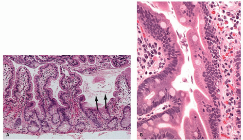
Figure 3-8. Intestinal giardiasis in a patient with common variable hypogammaglobulinemia. A: The jejunal mucosa is unremarkable. However, within the intestinal mucus are numerous giardial parasites (arrows). B: Detail of crypts showing numerous Giardia in the lumen, some probably attached to the epithelium.

Intrinsic and Associated Morphologic Changes Although the mucosal injury in the immunodeficient patient is frequently the result of superimposed infection, in certain disorders, there may be an intrinsic change independent of any recognized infection.12 For example, a case of IgA deficiency has been described, which had a severe “flat” mucosal lesion similar to that seen in celiac sprue, but the patient did not respond to a gluten-free diet. This patient was found to have an IgG antiepithelial cell antibody.86 In the stomach, pernicious anemia with gastric atrophy is found in some late-onset immunodeficiency syndromes. It occurs in younger patients (earlier than the usual 40-60 years
of age), lacks plasma cells in the atrophic mucosa, and lacks antibodies to intrinsic factor and parietal cells.76 Nodular lymphoid hyperplasia may be found in patients with CVID syndromes and selective IgA deficiency (Fig. 3-15).


Table 3-3 Incidence of Neoplasia in Immunodeficiency Disorders


Wiskott-Aldrich syndrome


15%


Ataxia telangiectasia


12%


Adult-onset common variable disease


10%


Isolated IgA deficiency


10%


Selective IgA deficiency


1%-3%


Childhood onset common variable disease


1%-3%


Severe combined immunodeficiency


1%-3%


Neoplasms The risk of neoplasia developing in immunodeficiency disorders is very high. In the primary immunodeficiency disorders, it has been estimated to be as high at 10,000 times that in the general age-matched population.87 The GI tract is frequently involved in neoplasia occurring roughly 10 to 200 times that of the general age-matched population (Table 3-3).68, 87, 88 The incidence of neoplasia varies with each type of immunodeficiency. The majority of malignancies are of lymphoid or hematopoietic origin (80%), the remainder being epithelial and smooth muscle.89 However, the type of malignancy varies with the immunologic disorder.87 Thus, in Wiskott-Aldrich syndrome, myelogenous leukemia is most common, whereas in common variable and selective IgA deficiency, epithelial tumors and lymphomas occur in approximately equal numbers. The types of neoplasms also vary with age and sex.88 For example, children with immunodeficiency usually develop non-Hodgkin’s lymphomas, whereas in adults, carcinomas and non-Hodgkin’s lymphomas are found equally often. In ataxia telangiectasia, females develop epithelial malignancies more frequently than males.

Although neoplasms in the immunodeficiency disorders involve most tissues, primary tumors of the GI tract occur fairly frequently, especially in common variable deficiency.76, 88 The overall incidence of tumors in one study of CVID was 25% of patients. Approximately half of these tumors occurred in the GI tract and consisted mainly of carcinoma of the stomach and colon or lymphoma (commonly associated with diffuse nodular lymphoid hyperplasia).87 An increased risk for neoplasia is also found in patients immunosuppressed for transplantation, or following chemotherapy for cancer.87 The risk of neoplasia in transplant patients is approximately 100 times greater than that observed in the general population in the same age range.87 In the transplant setting, there is about an equal incidence of developing lymphoid hyperplasia and lymphoma (posttransplant lymphoproliferative disorder, see “Lymphoma” section) and epithelial tumors, and the bowel is involved in approximately 12% of cases.87 Finally, GI tumors, primarily lymphoma, Kaposi’s sarcoma, and smooth muscle tumors,89 can be seen in untreated AIDS patients.


PRIMARY IMMUNODEFICIENCY DISORDERS

GI diseases due to infectious, inflammatory, and malignant disorders occur in a high proportion of patients with primary immunodeficiency disorders (Table 3-3).67, 74 The functional interaction of B and T cells and monocytes makes the division of immunodeficiency disorders somewhat artificial. Nevertheless, usually either antibody defects or cell-mediated abnormalities predominate, allowing a practical subdivision.15, 68, 69 This is the basis of the World Health Organization (WHO) classification of immunodeficiency disorders (Table 3-4). In addition, the WHO has included two other categories. One is characterized by the association of nonimmunologic defects. The other is characterized by a primary disorder of phagocytic function (chronic granulomatous disorder).


Predominant Antibody Defects

Common variable hypogammaglobulinemia (Late-onset acquired hypogammaglobulinemia, common variable immunodeficiency [CVID], Bruton’s X-linked agammaglobulinemia [XLAG])90, 91, 92

Pathogenesis and Clinical Features Common variable hypogammaglobulinemia, which is a heterogeneous group of immunologic disorders, is the second most common primary immunodeficiency disorder, affecting between 1/30,000 and 1/100,000 patients.93, 94 Three major immunologic abnormalities have been described to date, namely, an intrinsic B-cell defect, an immunoregulatory T-cell abnormality, and, rarely, the production of autoantibodies to T and B cells, all of which lead to impairment of immunoglobulin secretion.66, 67, 95, 96 A recent study found that naive B cells from patients with CVID express much lower levels of CD70 and CD86, molecules that are critical for T- and B-cell interactions.94 Hence, the T-cell abnormalities found in some of these patients may be secondary to an underlying B-cell defect.

In addition, this disorder is associated with celiac disease and occurs most commonly in the second to fifth decades.78, 97, 98 It is often familial and sometimes autosomal dominant or recessive modes of
inheritance can be discerned.76 Patients have panhypogammaglobulinemia and tend to present in one of two ways: either with recurrent infections, especially of the respiratory tract, or with chronic diarrhea and malabsorption.10, 74, 98 The intestinal symptoms are frequently due to giardiasis, although other parasites such as Cryptosporidium and strongyloidiasis as well as fungal overgrowth may also be responsible for the diarrhea and malabsorption.68, 69, 76, 80, 99 Other less common causes for chronic diarrhea are bacterial infections such as Campylobacter jejuni.100 These infections may sometimes be pathologically indistinguishable
from ulcerative colitis or result in mucosal fissuring and necrosis resembling Crohn’s disease.92, 101, 102 A third of these patients develop pernicious anemia and they may also develop a variety of other maladies, such as thyrotoxicosis, myxedema, arthritis, keratoconjunctivitis, splenomegaly, sarcoidosis, and amyloidosis secondary to infection.103 Most important, however, is the increased risk of GI tumors, especially gastric carcinoma in adults,88, 104 although lymphoma associated with nodular lymphoid hyperplasia as well as T-cell lymphoma unassociated with EBV viral infection have also been found.105, 106


Table 3-4 GI Manifestations of Primary Immunodeficiency Syndromes


TYPE


POSTULATED DEFECT


MAIN GI FEATURES


A. Predominant Antibody Defects


1.


Common variable immunodeficiency


  1. Predominant B-cell defect.


  2. Immunoregulatory T-cell disorder.


  3. Autoantibodies to B and T cells.


Intestinal infections due to Campylobacter, Salmonella, and Strongyloides. Spruelike syndrome due to Giardia or bacterial overgrowth. Pernicious anemia, nodular lymphoid hyperplasia, nongranulomatous ulcerative jejunoileitis, and GI carcinoma and lymphoma.


2.


Selective IgA deficiency


Defective IgA B-cell maturation.


Malabsorption due to bacterial overgrowth or parasite infestation, especially Giardia. Nodular lymphoid hyperplasia. Increased incidence of pernicious anemia, celiac disease, Crohn’s disease, and ulcerative colitis.


3.


Selective deficiency of other immunoglobulin isotypes, IgM, IgE, and IgD


Differentiation defect of IgM B cell to isotype-specific plasma cells.


Association with celiac disease, ulcerative colitis, Crohn’s disease, and Whipple’s disease.


4.


Infantile X-linked agammaglobulinemia (Bruton’s agammaglobulinemia)


Intrinsic defect of pre-B-cell to B-cell differentiation.


Malabsorption and chronic diarrhea due to bacterial overgrowth and persistent GI infections due to virus, Campylobacter fetus, Salmonella, and Giardia. Malignancy.


5.


X-linked immunodeficiency with increased IgM and IgD


Defect in isotype switch prevents normal maturation of IgM B cells to IgG, IgA, and IgE cells.


Diarrhea due to Candida infections and cryptosporidiosis. Malignancy.


6.


Transient hypogammaglobulinemia of infancy


Delayed B-cell maturation? due to defect in T-cell-dependent stimulation.


Malabsorption and diarrhea due to bacterial overgrowth, Campylobacter fetus and rotavirus infections, and Giardia infestations. Malignancy.


7.


Kappa chain deficiency


Unknown.


Achlorhydria, malabsorption, and diarrhea. Cystic fibrosis.


8.


Immunodeficiency with thymoma


Deficiency of pre-B cells? Defective development of stem cells.


Salmonella infections.


9.


Secretory component deficiency


Diarrhea, Candida infections.


B. Predominant Defects of Cell-mediated Immunity


1.


Severe combined immunodeficiency disease (includes Nezelof’s syndrome)


  1. Lymphoid maturation defect.


  2. T-cell defect due to adenosine deaminase deficiency.


  3. Purine nucleoside phosphorylase deficiency


Malabsorption and intractable diarrhea, repeated GI viral and fungal infections (herpes, varicella, rotavirus, and Candida).


2.


Chronic mucocutaneous candiasis


Uncertain, may be secondary to drugs.


Mouth ulcers, esophagitis, esophageal stricture, and diarrhea


C. Immunodeficiency Associated with Other Defects


1.


Wiskott-Aldrich syndrome


Cell membrane defect affecting all hemopoietic stem cell derivatives.


Recurrent bloody diarrhea and malabsorption.


2.


DiGeorge’s syndrome (congenital thymic aplasia)


Abnormal thymic development with resultant T-cell defects.


Malabsorption, intractable diarrhea, and severe malnutrition.


3.


Ataxia telangiectasia


Defective T-cell maturation.


Nodular lymphoid hyperplasia, vitamin B12 malabsorption.


4.


Transcobalamine deficiency


Autosomal recessive transcobalamine II deficiency.


Intractable diarrhea.


D. Phagocyte Dysfunction


Chronic granulomatous disease


Macrophages unable to kill phagocytosed organisms.


GI obstruction, malabsorption and diarrhea.


Histology The pathologic changes are found most commonly in the small intestine but may occur throughout the entire GI tract. There is a wide range of histologic findings including nonspecific mucosal lesions, features resembling acute graft versus host disease (GVHD), atrophic gastritis, hypogamma-globulinemic sprue-like features with mild to severe villous atrophy, granulomatous changes resembling Crohn’s disease, histologic features resembling Whipple’s, and nongranulomatous ulcerative jejunoileitis. In addition, nodular lymphoid hyperplasia and malignancy, especially gastric carcinoma, can occur.78, 87, 88, 92, 93, 102, 104 The pathologic findings do not always correlate with the severity of clinical findings.

STOMACH. An atrophic gastritis similar to that of pernicious anemia but lacking the plasmacytic infiltrate is seen in some cases of CVID disease. In contrast to the usual cases of pernicious anemia, these patients do not have autoantibodies and serum gastrin levels are not elevated.107, 108 Helicobacter pylori appears to be an important cofactor in causing multifocal atrophic gastritis in CVID patients and probably helps explain their high incidence of gastric carcinoma.104

Only gold members can continue reading. Log In or Register to continue

Stay updated, free articles. Join our Telegram channel

Jul 20, 2016 | Posted by in GASTROENTEROLOGY | Comments Off on Immunodeficiency Disorders

Full access? Get Clinical Tree

Get Clinical Tree app for offline access