Diagnostic accuracy
Radiation exposure
Cost
Non-contrast computed tomography
++++
+
+
Low dose NCCT
+++
++
+
Ultrasound
++
++++
++
Plain abdominal radiography (KUB)
+
+++
++++
KUB with tomograms
++
+
+++
Digital tomosynthesis
++
+++
+++
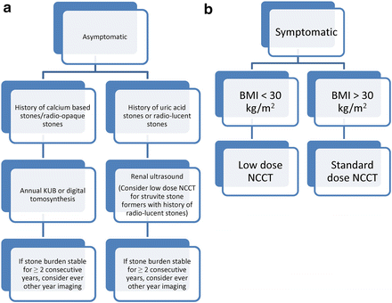
Fig. 14.1.
Imaging of the recurrent stone former. (a) imaging for the asymptomatic patient (b) imaging for the symptomatic patient
References
1.
Pearle MS, et al. Medical management of kidney stones: AUA guideline. J Urol. 2014;192(2):316–24.PubMedCrossRef
Related posts:


Stay updated, free articles. Join our Telegram channel

Full access? Get Clinical Tree


