, Ivan Damjanov1 and Ryan M. Taylor2
(1)
University of Kansas School of Medicine, University of Kansas, Kansas City, KS, USA
(2)
Division of Gastroenterology and Hepatology, University of Kansas School of Medicine, Kansas City, KS, USA
Hepatocellular Changes
Ballooned hepatocyte (Fig. 1)
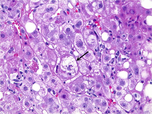
Fig. 1.
Ballooned hepatocyte. A ballooned hepatocyte (arrow) is a hepatocyte that is 3–4 times the size of neighboring hepatocytes and has cleared out cytoplasm with clumpy intermediate filaments
3–4 times the size of neighboring hepatocytes.
Cleared out cytoplasm and strands of clumpy intermediate filaments (some of which are prominent enough to be classified as Mallory-Denk bodies).
Non-specific finding that is usually associated with steatohepatitis or cholestasis but can be seen in a wide variety of changes related to oxidative stress.
Can be easily seen on a trichrome stained section and are often the cells that are associated with pericellular/sinusoidal fibrosis.
“Dead red” (Fig. 2)

Fig. 2.
Apoptotic hepatocyte. Apoptotic hepatocyte or “dead red” (arrow) is a brightly eosinophilic hepatocyte
Single dead hepatocyte in the lobule with eosinophilic cytoplasm.
No nucleus or a very pyknotic (dark) nucleus.
This cell goes by many names including apoptotic hepatocyte/hepatocyte apoptosis, acidophil body, Councilman body, spotty necrosis, and dead hepatocytes.
Feathery degeneration/cholate stasis (Fig. 3)
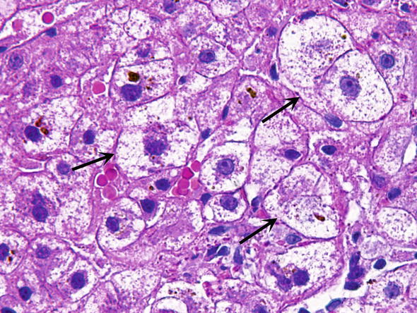
Fig. 3.
Feathery degeneration. Hepatocytes with feathery degeneration (arrow) are often seen in association with cholestasis
Pale hepatocytes seen in intrahepatic cholestasis.
Related to the detergent type action of bile acids on the hepatocyte.
More pronounced in zone 1 or the periportal hepatocytes—in contrast to the ballooning degeneration typically associated with steatohepatitis, which is usually most prominent in the centrilobular zone.
Hemosiderosis
Brown, granular refractile cytoplasmic deposits on H&E stained sections (Fig. 4a).
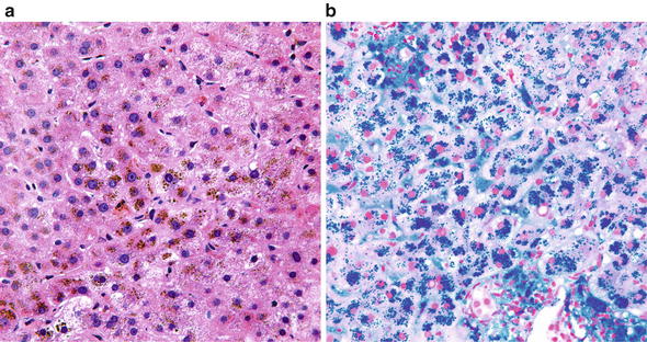
Fig. 4.
Hemosiderosis. (a) In routine H&E stained sections, iron appears as brown granular pigment. (b) Prussian blue stain performed according to Perl makes the hemosiderin granules appear blue
Bright blue chunky deposits on Perl Prussian blue stain (Fig. 4b).
Hepatocyte drop out/confluent necrosis (Fig. 5)
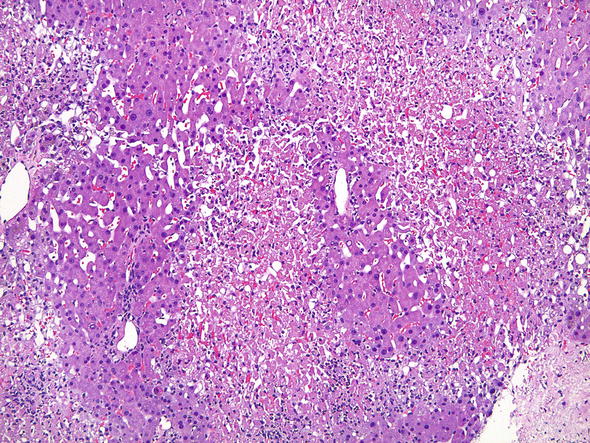
Fig. 5.
Confluent necrosis. Confluent necrosis is characterized by large zones of hepatocyte death
Hepatocyte drop out and confluent necrosis are synonymous and refer to ZONES of hepatocyte death; in contrast to individual cell apoptosis aka “dead reds”.
Pattern of injury that can be seen in some presentations of acute hepatitis such as autoimmune hepatitis or plasma cell rich hepatitis in transplant grafts.
Severe forms of this pattern of injury are referred to as panacinar necrosis or submassive necrosis.
Mallory-Denk bodies (Fig. 6)
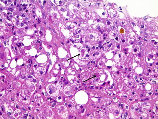
Fig. 6.
Mallory-Denk bodies. Mallory-Denk bodies (arrows) appear as thick ropy eosinophilic clumps within a ballooned hepatocyte
Ubiquitinated cytokeratin that appears as bright eosinophilic ropy clumps aggregated within the hepatocyte cytoplasm.
Often associated with steatohepatitis and within ballooned hepatocytes.
Steatosis (Fig. 7)
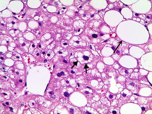
Fig. 7.
Steatosis. Steatosis most often exists as small droplet (closed arrows) and large droplet (open arrow) macrovesicular steatosis
Fat accumulation in the liver.
Vast majority of steatosis in the liver exists as macrovesicular steatosis—both small droplet and large droplet.
Large droplet macrovesicular steatosis—a large single droplet of lipid that completely displaces the nucleus off to the side of the cell.
Small droplet macrovesicular steatosis—the nucleus is still centrally located and there are multiple vacuoles in the cytoplasm, some of which indent the nucleus.
Most biopsies which show steatosis show a combination of both small and large droplet steatosis—this pattern of steatosis is common in alcoholic and non-alcoholic steatohepatitis and is also associated with drug-induced liver injury.
Steatosis does not have associated inflammation. Steatohepatitis is the combination of both steatosis and inflammation.
Microvesicular steatosis is actually quite rare and refers to tiny droplets filling the entire cytoplasm and is associated with disease not seen very often biopsied, such as fatty liver disease of pregnancy and valproic acid toxicity (Fig. 8).
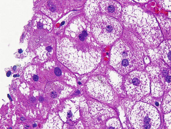
Fig. 8.
Microvesicular steatosis. Microvesicular steatosis appears as tiny innumerable droplets within the cytoplasm that do not displace the nucleus
Steatohepatitis (Fig. 9)
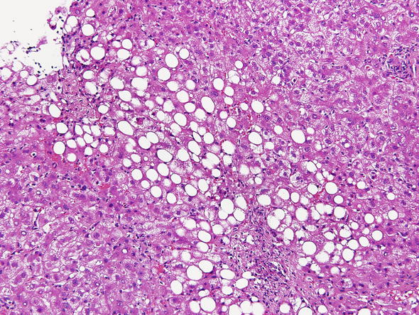
Fig. 9.
Steatohepatitis. Steatohepatitis is the combination of steatosis, lobular inflammation, and ballooned hepatocytes
Steatohepatitis is the combination of steatosis and lobular inflammation, usually with ballooned hepatocytes as well.
Nonspecific pattern of injury seen most commonly with morbid obesity, diabetes, insulin resistance, and alcohol abuse. It can also be associated with nutritional causes, metabolic disorders or the result of certain medications.
Inflammation
Endotheliitis (Fig. 10)
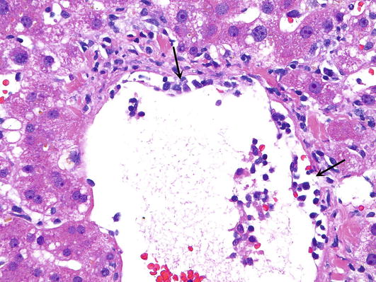
Fig. 10.
Endothellitis. Endotheliitis is characterized by inflammation of the endothelium. The arrow demarcates the basement membrane of the central vein which has been lifted due to inflammation
Lymphocytes beneath the portal and central vein endothelium.Related posts:

Stay updated, free articles. Join our Telegram channel

Full access? Get Clinical Tree


