Fig. 8.1
Flexible ureterorenoscopes available. Left are fiber optic ureteroscopes, and on the right are the digital ureteroscope. The Olympus V2 is coming soon and is not illustrated
In 2001, the DUR-8Elite by ACMI became the first new generation flexible ureteroscope that had two levers: primary down (180°), primary up (170°), and secondary down (130°). Following the DUR-8Elite, other manufacturers produced the newer generation ureteroscopes, though these scopes had a single lever (Table 8.1).
Company | Product | Imaging system | Ventral deflexion (°) | Dorsal deflexion (°) | Tip diameter (F) | Shaft diameter (F) | Proximal diameter (F) | French scale test |
|---|---|---|---|---|---|---|---|---|
Olympus Gyrus ACMI | DUR-8E | Optical | 270 | 270 | 8.7 | 9.4 | 10.1 | 10 |
DUR-8U | Optical | 270 | 270 | 8.6 | 9.36 | 10.1 | 10 | |
DUR-D | Digital | 250 | 250 | 8.7 | 9.3 | 10.1 | 11 | |
Olympus | URF-P5 | Optical | 275 | 180 | 5.3 | 8.4 | 8.4 | 10 |
URF-P6 | Optical | 275 | 275 | 4.9 | 7.95 | 7.95 | 10 | |
URF-V | Digital | 275 | 180 | 8.4 | 10.9 | 10.9 | 12 | |
URF-V2 | Digital | 275 | 275 | 8.4 | 8.4 | 8.4 | NA | |
Storz | Flex-X2 | Optical | 270 | 270 | 7.5 | 8.4 | 8.4 | 10 |
Flex-XC | Digital | 270 | 270 | 8.5 | 8.5 | 8.5 | 10 | |
Wolf | Cobraa | Optical | 270 | 270 | 6 | 9.9 | 10.3 | 11 |
Viper | Optical | 270 | 270 | 6 | 8.8 | 9 | 10 | |
Boa vision | Digital | 270 | 270 | 6 | 8.9 | 8.9 | NA | |
Cobra Visiona | Digital | 270 | 270 | 6 | 9.9 | 9.9 | NA | |
Stryker | Flex vision U-500 | Optical | 275 | 275 | 6.9 | 7.1 | 7.2 | 10 |
Lumenis | PolyScope | Optical | 180 | 0 | 9.6 | 8 | 8 | 10 |
Currently, with the arrival of the Olympus URF-P6 ureteroscope, each ureteroscope manufacturer produces a flexible ureteroscope with at least 270° of deflection in both directions. Moreover, some of the latest models are categorized as a “semi-flexible ureterorenoscope” (Fig. 8.2). A “semi-flexible ureterorenoscope” has more strength in the ureteroscope shaft and facilitates the ureteral ascension as well as allows for increased scope control once in the renal pelvis during flexible nephroscopy. Other recent flexible ureteroscopes also tend to have a smaller tip with each scope fitting inside the 10/12F UAS [7] (Table 8.1).
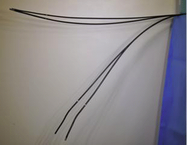
Fig. 8.2
The new ureteroscopes tend to be “semi-flexible.” The newer Olympus URF-V2 and URF-P6 are at the top of the picture, and older Olympus URF-V and URF-P5 are both at the bottom
All current flexible ureteroscopes have acceptable optical quality with a 0° optic (except for a 9° optic for the Gyrus ACMI DUR-8Elite) and a field of view of 80–88°. The instruments are fragile and require manipulation and use with caution during the surgery as well as during the sterilization process. The surgeon should never insert an instrument into the working channel with a bent distal tip in order to prevent damage. With recent technological advances, the new generation of ureteroscopes are more durable and the utilization time before requiring a repair has increased up to 76 h longer compared to older generation of ureteroscopes. Some authors have also described performing up to 100 interventions with the same flexible ureterorenoscope without requiring repair [9, 10].
Newer Generation Digital Flexible Ureterorenoscopes
Since 2006, the digital optics technology has represented the next step in the development of endoscopy with a distal sensor. Studies have showed that the better image quality provides a higher precision for diagnosis, treatment, and a shorter procedure time (Fig. 8.3) [11].
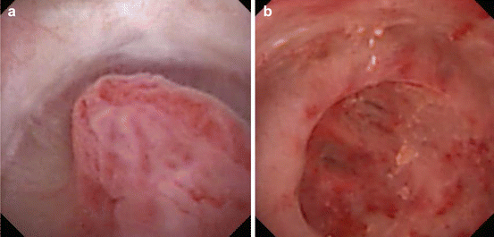
Fig. 8.3
Improved image quality of digital ureteroscopes: (a) Angioma of the renal papilla and (b) Papillary necrosis diagnosed in the context of hematuria
The first ureteroscopes to use digital technology include was Gyrus-ACMI DUR-D Invisio, Karl Storz with the Flex-XC, the Olympus the URF-V and URF-V2, and the Richard Wolf Boa and Cobra Vision. These new ureteroscopes integrate a digital camera and light source in a single plug and play device. The principal disadvantages with the digital generation are that the distal tip diameter are slightly greater than the equivalent standard fiber optic ureteroscopes (Fig. 8.4). Nevertheless, the digital generation is now considered the gold standard in term of image quality.
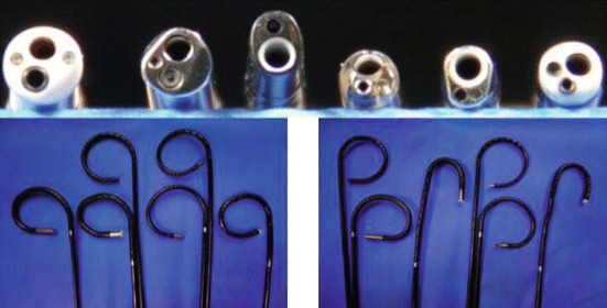
Fig. 8.4
Distal tips and active deflections for flexible ureteroscopes. Left is downward deflection and right is upward deflection. From left to right: Olympus URF-V, URF-V2, Storz Flex-XC, Olympus URF-P6, Storz Flex-X2, Olympus URF-P5
All digital ureteroscopes by Karl Storz, Gyrus-ACMI and Richard Wolf use a Complementary Metal Oxide Semiconductor (CMOS) imaging sensor as a digital camera and a light-emitting diode (LED) as the light source. CMOS is a generic term for the process used to produce these image sensors, along with a multitude of other semiconductor items such as computer RAM processors like those from Intel and other manufacturers. CMOS image sensors can be made in the same fabrication as these other items, with the same equipment.
Olympus has introduced the URF-V incorporating a charge-coupled device sensor (CCD). Interline Transfer CCD consists of many Metal Oxide Semiconductor (MOS) capacitors arranged in a pattern that can capture and convert light photons into electrical charge, storing this charge before transferring it for processing by supporting chips. When recording color information, a color filter is placed over each light receptor making it sensitive to only one light color. This gives a value for one color at each pixel, and the surrounding pixels can provide eight more values, four each for the two remaining colors from which they may be interpolated from the original pixel. After the exposure is complete, the charge is transferred row by row into a read-out register, and from there to an output amplifier, analog/digital converters and on for processing. This row-by-row processing of the CCD’s light data is where the sensor gets register and the rows behind it are each shifted one row closer to the register. After being “read-out,” the charge is released, and the register is empty again for the next charge. CCDs rapidly attain high image quality but require a manufacturing process which is different from that used for manufacturing other computer chips such as processors and RAM. This means that specialized CCD fabrications have to be constructed, and they cannot be used for making other components, thereby making CCDs more expensive.
Moreover, the Olympus URF-V incorporates another new technology: the narrow band imaging (NBI) function. NBI is an optical image enhancement which narrows the bandwidth of the light production of the Olympus Exera II system. This narrow band of light is greatly absorbed by haemoglobin and penetrates only the surface of tissue increasing the visibility of capillaries and other delicate tissue surface structures by enhancing the contrast between the two. As a result, under NBI, capillaries on the surface are displayed in brown and veins in the sub-surface are displayed in cyan on the operating monitor. This technology is principally used in the detection or recurrence of urothelial tumors of the bladder or upper urinary tract. One of the current authors (OT) has demonstrated that NBI has the potential to improve the detection/recurrence of upper tract urothelial tumors by 22.7 % comparing to white light (Fig. 8.5) [12].

Fig. 8.5
(a) Upper tract urothelial tumor visualized with the white light. (b) Tumor visualized with the Narrow Band Imaging function
Disposable Flexible Ureteroscope
In 2009, the PolyScope by Lumenis became available. This is a disposable, modular flexible ureteroscope with a 70 cm working length and a distal tip size of 9.6F. It has a single-side active deflection of 180°, and the working channel is 3.6F [13]. Currently, more studies are needed to demonstrate the efficiency and cost-effectiveness of this device though it does appear to be more difficult to manipulate than the usual flexible ureterorenoscopes.
How to Easily Traverse the Ureter
Getting ureteral access and reaching the proximal ureter or the renal pelvis can sometimes be challenging. Thus knowing alternative access methods to assist the endourologist is essential.
Ureteral Cannulation
Guidewires are well known to be essential during F-URS. Their use can be helpful for UAS placement or for the ureteroscope ascending a tortuous ureter and remain the best security during the surgery if a ureteral stent insertion is rapidly required. There are many types of guidewires and the description of guidewires is further discussed in Chap. 11. The authors strongly recommend using the stiff hydrophilic wires, where the first 3–5 cm are completely floppy hydrophilic to be less traumatic and the shaft of the wire is stiff and hydrophobic. Standard floppy tip polytetrafluoroethylene (PTFE) could also be used, but hydrophilic wire have been shown to be superior over the standard PTFE in gaining access to the renal pelvis when passing by a ureteral stone [14, 15]. The tip can be straight, angled or “J” tip and the surgeon, based on his/her own experience, will need to decide when he/she should use an angled one over a straight wire (Fig. 8.6).
Fig. 8.6
Different guidewire tips
When two wires are used and the flexible ureteroscope is placed over the working wire, you should use a hydrophilic wire to protect the working channel of the scope. Going up into the ureter directly over the wire facilitates the scope placement into renal cavities. We strongly recommend using a safety wire even if the literature has demonstrated the feasibility of F-URS without a safety wire [16].
If difficulties are encountered in placing the guidewire during cystoscopy, a 5–7Fr open-end ureteral catheter can be used to accommodate the positioning of the guidewire into the ureter. The catheter is made of polyurethane and retrograde pyelography can also be performed in order to further delineate ureteral anatomy (Fig. 8.7).
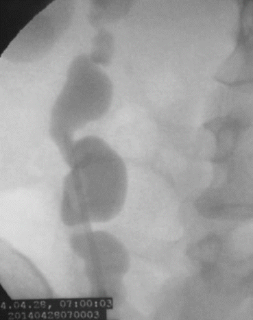
Fig. 8.7
Retrograde pyelography of a patient with an ileal ureter
Another use for the ureteral catheter is in the setting of a false passage caused during ureteral orifice cannulation. In this situation, a whistle tip ureteral catheter with a hydrophilic angled-tip guidewire can be used to help to find the correct passage into the distal ureter (Fig. 8.8). Following a right sided creation of a false passage, the correct way into the ureter is found by orienting the open-end and the angle-tip guidewire at 11 o’clock at the ureteral orifice. On the left side, you will have to orient the open ended catheter and angled guide wire to 2 o’clock.
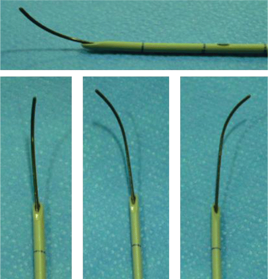
Fig. 8.8
Whistle tip ureteral catheter with angle-tip guidewire showed in different positions
Ureteral Access Sheath
The ureteral access sheath (UAS) was first developed in the 1970s to help gain ureteral access during ureteroscopy. One decade later, a peel-away sheath was introduced. The UAS has increased in use with the rapidly growing rate of retrograde intrarenal surgery (RIRS) during the past few years. Using a UAS is not mandatory during RIRS, but it does allow easy repeat passage into and out of the ureter with the flexible ureteroscope. Moreover, it dilates the ureter and improves visualization by facilitating the return of irrigation. It also allows one to perform the surgery with lower intrarenal pressure thus reducing the risk of intrarenal reflux. It encourages the passage of small stone fragments and protects the flexible ureteroscope and gives more stability to the scope that permits optimal control during instrumentation into the renal pelvis [17].
Related posts:
 Radiation Exposure to the Patient and the Urologist
Shock Wave Lithotripsy for the Treatment of Ureteral Stones
Selecting the Appropriate Treatment Modality for Ureteral Calculi
Complications in the Treatment of Ureteral Stones: Prevention and Management
Radiation Exposure to the Patient and the Urologist
Shock Wave Lithotripsy for the Treatment of Ureteral Stones
Selecting the Appropriate Treatment Modality for Ureteral Calculi
Complications in the Treatment of Ureteral Stones: Prevention and Management
 Semirigid Ureteroscopy for Ureteral Calculi
 Tips and Tricks in the Treatment of Ureteral Stones
Stay updated, free articles. Join our Telegram channel

Full access? Get Clinical Tree


