Indwelling preoperative stent
No preoperative stent
Male
13/15 F or 14/16 F, 45 cm
11/13 F or 12/14 F, 45 cm
Female
13/15 F or 14/16 F, 35 cm
11/13 F or 12/14 F, 35 cm
The correct placement of the UAS allows maintaining low intrarenal pressures during flexible ureterorenoscopy. Maintaining intrarenal pressure below 40 ccH2O during flexible ureteroscopy prevents pyelovenous and/or pyelolymphatic backflow during the procedure. Rehman et al., studied intrarenal pressures in cadaveric kidneys using 10/12, 12/14, and 14/16 F UAS, and compared this to controls without a UAS. With the ureteroscope in place, irrigation was run at pressures of 50, 100, and 200 cmH2O. Without a UAS, intrarenal pressures rose to 52–59 cmH2O, high enough to cause pyelovenous and pyelolymphatic backflow. With the use of a UAS lower pressures were observed. The 10/12 F access resulted in pressures of 22–29 cmH2O, while the larger 12/14 and 14/16 F sheaths had lower pressures with measurements of 17.5–19.5 and 14.5–19.5 mmH2O, respectively [7]. This suggests, at least in terms of intrarenal pressure management, that a 12/14 F UAS strikes the best balance between size of the sheath and effectiveness.
During treatment of upper tract urothelial cell carcinoma, a properly selected and deployed UAS may aid in preventing extravasation of tumor cells by maintaining low intrarenal pressures. The sheath may also reduce the exposure of the lower portions of the ureter to tumor cells as it effectively covers the urothelium and promotes the flow of irrigation out through the sheath directly rather than around it.
With a UAS in position, the surgeon is able to introduce and remove the ureteroscope at will during the procedure. When a tumor is being investigated, multiple biopsies with larger instruments such as a back-loading biopsy forceps can be performed. However, the greatest advantage may be in the treatment of renal calculi. After laser lithotripsy of an upper ureteral or renal stone, the surgeon is able to basket extract the fragments through the UAS in an attempt to render the patient stone-free. The size of the fragments removed is limited by the diameter of the access sheath. The access sheath protects the ureter from the inadvertent removal of fragments that are too large and that could get trapped in the ureter, or worse, avulse the ureter if a sheath were not used. If fragments are attempted to be removed that are too large for the sheath, they are then further reduced in size with laser lithotripsy before further removal is attempted.
Removal of the fragments has several potential benefits for the patient. When the patient passes the fragments they may experience episodes of renal colic. Worse, if the fragments became obstructed, further intervention may be required including possible stent placement or further lithotripsy. If the fragments fail to pass and they remain in the kidney, then they could become the nidus for future stone formation [8, 9]. In a nonrandomized study, L’Esperance et al. demonstrated that the use of a UAS resulted in an 12% higher stone-free rate as compared to procedure where an access sheath was not used (79% vs. 67% SFR, respectively) [10].
Kourambas et al. performed a study that randomized 59 patients who were scheduled to undergo ureteroscopy into two groups—one with a UAS and one without. The majority (85%) of the procedures were performed for stone disease. They determined that there was a statistically significant reduction in the mean operating time by 10.5 min when an access sheath was employed during the procedure. Furthermore, despite the cost of the access sheath, the overall cost per patient was reduced by $350 in the cases where the sheath was used. Contrary to L’Esperance’s data, this study showed no difference in the stone-free rate between the two groups. This may be due to the lack of stone basketing in the access sheath arm. Short-term follow-up did not reveal an increased rate of ureteral strictures in the access sheath arm of the study [6].
Ischemia
A concern with the placement of a UAS has been whether ischemia to the ureter can occur. In an animal study, Lallas et al. studied the potential for ischemia in porcine ureters after the placement of an access sheath using laser Doppler flowmeter. Access sheaths of 10/12, 12/14, and 14/16 F were placed and measurements were taken at 5 min intervals from the proximal ureters. Blood flow was reduced in all ureters at the first 5 min interval, but the greatest reduction in blood flow occurred with the larger 12/14 and 14/16 F sheaths, where flow decreased greater than 50% by 15 min. However, blood flow subsequently increased to 70% of baseline levels by the 70 min mark. Histologic exam of the ureters taken at 72 h after the procedure showed no evidence of ischemia or necrosis [11]. This suggests that despite the initial decrease in blood flow, the impact is transient and the reduction is not enough to induce ischemic necrosis.
Stricture Risks
The transitory reduction in ureteral blood flow raises the question as to whether there could be an increased risk of stricture formation in patients who undergo ureteroscopy with a UAS. To date, no randomized data has shown this to be a risk. In a nonrandomized study, Delvecchio et al. reviewed the records of 150 patients who underwent ureteroscopy with the use of a UAS. Sixty-two of these patients had follow-up in excess of 3 months. In this subgroup, only one patient developed a stricture suggesting that the overall stricture rate is similar to previously published stricture rates after ureteroscopy without a sheath [12, 13].
Stent or No Stent
The debate whether to place a stent after ureteroscopy has continued over the last decade. Several studies have shown that in uncomplicated ureteroscopy that routine stenting is not indicated and that unstented patients had decreased pain, narcotic use, and morbidity [14, 15]. However, this work was done prior to the widespread use of UASs and these studies do not discuss the use of a UAS. The placement of a UAS can result in some minor trauma to the ureter during insertion as well as transient ischemia [11]. Rapaport et al. reported in a nonrandomized series of patients that 37% of patient who underwent a ureteroscopy with a UAS returned for an unscheduled emergency visit in the postoperative period vs. 14% of patients who underwent ureteroscopy without an access sheath. They recommended that a stent be placed after all ureteroscopy cases involving a UAS [16]. It is not entirely clear what led to the increased rate of ER visits in the group that had a UAS placed during surgery, but transient colic for spasm or edema of the ureter is a possible explanation.
Reducing Scope Damage
The limited durability and high cost of repair of modern flexible ureteroscopes is a financial hurdle that can be difficult to overcome for some centers wishing to adopt the technology [17]. In a nonrandomized study using historical controls, Pietrow et al. reported that when UAS were used, they were able to double the number of procedures before repairs to their endoscopes were needed as compared to when no access sheath was used (27.5 uses) [18]. Several factors could be contributing to this. When a UAS is not used, the flexible ureteroscope is usually passed up over a guide wire (railroad technique). This can result in damage to the endoscope channel from the wire, especially with wires that have a rigid back end. Small perforations to the channel of the ureteroscope will lead to leaks that will further damage the endoscope. Small flaps in the channel can also be created that may make the passage of instruments difficult. Furthermore, when the resistance is encountered during the advancement of the ureteroscope, the scope can buckle from the force. Given the delicate design of these scopes, this may also lead to further damage. Buckling of the scope is prevented when it is advanced through a UAS.
Properties of Different Access Sheaths
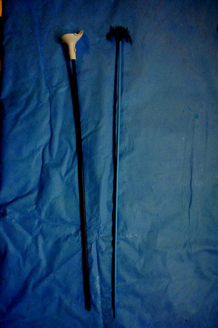
There are a wide variety of commercially available UASs that differ in their physical properties. The properties of the sheath may impact how clinically effective they are. Monga et al. evaluated in a bench study eight UAS to determine their resistance to buckling and kinking as well as lubriciousness (coefficient of friction). They determined that the Applied Forte XE (Applied Medical, Rancho Santa Margarita, CA) and Cook Flexor sheaths (Cook Medical, Bloomington, IN) were the most resistant to kinking as well as most lubricious (Fig. 24.1). The Cook Flexor also had the greatest resistance to buckling [19]. By resisting kinking, the lumen of the access sheath may be less likely to collapse during a procedure. Having a lower coefficient of friction, may allow the access sheath to slide up the ureter more easily but could also make it more susceptible to slipping backwards during a procedure. Resistance to buckling may allow the sheath to pass easier when some resistance is encountered, such as at the ureteral orifice or pelvic brim.


Fig. 24.1
UAS showing inner obturator and outer sheath
Using this data, a prospective randomized trial was performed comparing these two sheaths in a clinical study. Fifty-four patients were randomized to flexible ureteroscopy procedures with either a 12/15 F Applied Access Forte XE or 12/14 F Cook Flexor. Both of these sheaths are reinforced with an embedded coil system and feature a hydrophilic coating. The overall device failure rate for the Applied Access Forte XE was 44% vs. no failures for the Cook Flexor. Buckling (25%), kinking (25%), and difficulty passing instruments (13%) were the causes of failure for the Applied sheath. Of note, in procedures where the Applied sheath failed, the Cook Flexor sheath was then attempted to be placed. In all cases the Cook Flexor was successfully placed. The Cook Flexor was also scored higher subjectively by the surgeon in terms of ease of placement, instrument passage, and ease of stone extraction. The authors speculated that the larger outer diameter of the Applied sheath (15 F vs. 14 F) and the length and configuration of the tip of the sheath may have been a factor in the outcome of the study [20].
A follow-up study evaluated “next-generation” UASs in terms of buckling and kinking. Sheaths evaluated including the Cook Flexor (12/14 F, 35 cm), ACMI UroPass (12/14 F, 38 cm) (Gyrus ACMI, Southborough, MA), Bard Aquaguide (11/13 F, 35 cm) (Bard Medical, Covington, GA), and Boston Scientific Navigator (11/13 and 13/15 F, 36 cm) (Boston Scientific, Natick, MA). The Cook Flexor was again the most resistant to buckling requiring 5.1 N to buckle, followed by the ACMI at 3.2 N, the Bsci-13/15 at 2.9 N, the Bard at 2.8 N, and the Bsci-11/13 at 2.0 N. The smaller diameter of the Bard and BSci 11/13 appear to make them more prone to buckling. The Bard sheath was the most prone to kinking, requiring only 9 N/mm to do so. The BSci 13/15 kinked at 30 N/mm, the BSci 11/13 at 41 N/mm, the Cook at 42 N/mm, and the least likely to kink was the ACMI at 83 N/mm. All sheaths tested had hydrophilic coatings and showed similar coefficient of frictions [21].
Our own clinical experience has been that kinking of the sheath during a procedure is rarely seen with a modern coil reinforced access sheath UAS but buckling can be encountered with advancement of the sheath. Passing the UAS over an Amplatz superstiff guide wire may reduce the incidence of buckling, but care must be taken to not exert too high of a force in order to avoid trauma to the ureter. Study has shown that experienced urologists use up to 6.6 N when placing an access sheath [22]. While the exact force needed to perforate a ureter while advancing an access sheath over a guide wire is variable depending on both the type of UAS used as well as individual patient factors, a CT-1 needle requires 4.7 N of force to perforate a human ureter [23].
UAS and Flexible Ureteroscopes Facilitating Other Procedures
The use of a flexible ureteroscope combined with a UAS can be advantageous during percutaneous stone procedures. By placing a UAS in a retrograde fashion at the start of a percutaneous nephrolithotomy (PCNL), the access sheath allows for small stone fragments to pass out through the sheath and not become lodged in the ureter. This also assists in maintaining low pressures within the renal collecting system during the procedure. A flexible ureteroscope can be advanced up into the kidney during the procedure to visualize the best calyx prior to percutaneous puncture. Contrast or air can be injected through the scope to facilitate puncture, but the tip of the scope can also serve as the fluoroscopic target. With the scope in position, the puncture of the needle is directly visualized as it enters into the collecting system. The guide wire can also be captured with a stone basket passed through the endoscope and withdrawn out through the UAS [24]. The flexible ureteroscope with laser lithotripsy and basket extraction can also be used to clear residual stone burden that could not be reached in an antegrade fashion during the PCNL procedure. This may reduce the need for multiple nephrostomy tracts in patients with large or complex stone burdens [25].
Radially Dilating Balloon
A novel UAS that incorporates a radially dilating balloon has been reported. The proposed advantages of a radially dilating balloon incorporated into the UAS are that it eliminates the shearing forces associated with axial dilation. This is a reported advantage to using balloon dilation vs. radial dilation during PCNL and may translate to similar findings with UAS placement [26]. Harper et al. reported their experience placing a balloon expandable ureteral access (BEUS) and compared it to placement of a conventional sheath (Cook Flexor, 12/14 F) in ten farm pigs. The BEUS is 9.5 F in nonexpanded form and after placement it expands to have an inner diameter of 12 F and an outer diameter of 14 F. The study demonstrated that the BEUS required less maximum (0.36 lb vs. 1.48 lb) and less mean force (0.11 lb vs. 0.49 lb) to place vs. the conventional UAS. The flow rate through the BEUS was also slightly improved over the conventional UAS (90.0 cc/min vs. 80.6 cc/min). Blinded reviewers also scored the BEUS superior for less total urothelial tear length (1.2 cm vs. 2.6 cm) and less damage to the ureter overall [27]. This type of access sheath design holds promise as an advancement over current designs. However, some changes to the technique of access sheath placement may be required. It would be important to ensure that there are no unrecognized strictures in the ureter prior to placement. If a tight stricture were to be encountered that the sheath could not dilate, it would require pulling the expanded sheath through the stricture. It is unclear as to how much force this would place on the ureter but the concern would be that enough force could be generated to risk of avulsion of the ureter. This force would be variable depending on both the location of the stricture and other patient factors. By performing a retrograde ureterogram prior to the placement of the sheath, one would be able to identify a previously unrecognized stricture and treat it appropriately prior to placement of the BEUS.
Stay updated, free articles. Join our Telegram channel

Full access? Get Clinical Tree


