, Adele Taibbi1 and Massimo Midiri1
(1)
Department of Radiology, University Hospital, Palermo, Italy
Diffuse fatty infiltration is a common imaging finding due to the abnormal accumulation of lipids within hepatocytes. Baseline US is typically the first imaging modality for the evaluation of hepatic steatosis, but causing a conspicuous increase in liver echogenicity and a marked ultrasound beam attenuation, it really hampers FLL detection and characterization. Moreover, geographic fatty changes of the liver can occur when the fat accumulation ability of hepatocytes or fat deposition becomes heterogeneous throughout the liver and are classified into four subtypes: (a) focal fatty change, (b) multifocal steatosis, (c) lobar or segmental steatosis, and (d) focal fatty sparing in fatty liver [1].
In particular, focal fatty change and focal fatty sparing can show a round, mass-like appearance on imaging, making difficult a correct differential diagnosis with malignant lesions in oncological patients. At this regard, CEUS has been reported to improve the characterization of focal hepatic lesions in patients with fatty liver and in diagnosing both focal fatty infiltration and sparing [2]. But there are different and controversial data in literature about the usefulness of CEUS in the screening and follow-up of oncological patients [3–9]. Anyway, WFUMB-EFSUMB guidelines recommend CEUS in the surveillance of oncological patients (if CEUS has been useful previously), for the evaluation of liver metastases in colorectal cancer after chemotherapy instead of unenhanced US, or for lesion(s) or suspected lesion(s) detected with US in patients with a known history of a malignancy as an alternative to CT or MRI [10–12].
4.1 Diffuse Fatty Liver
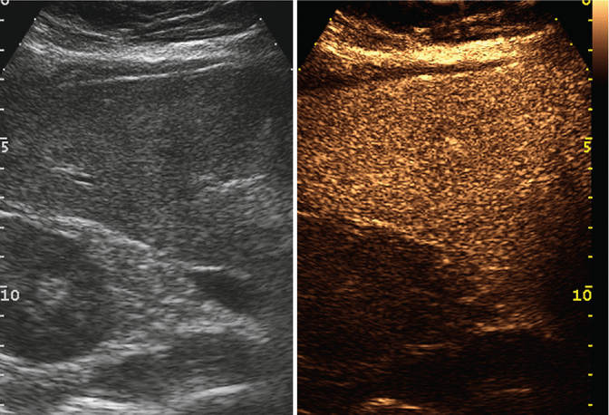
Fig. 4.1
Diffuse fatty change in a 65-year-old man. At unenhanced US (left image), diffuse fatty change is appreciable. At CEUS (right image), homogeneous contrast enhancement is evident in the extended portal-venous phase
4.2 Geographic Fatty Change
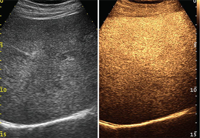
Fig. 4.2
Geographic fatty change in a 49-year-old woman. At unenhanced US (left image), diffuse inhomogeneous fatty infiltration is evident. At CEUS (right image), homogenous contrast enhancement is evident in the extended portal-venous phase
4.3 Focal Fatty Change
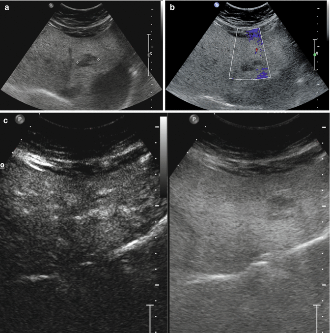
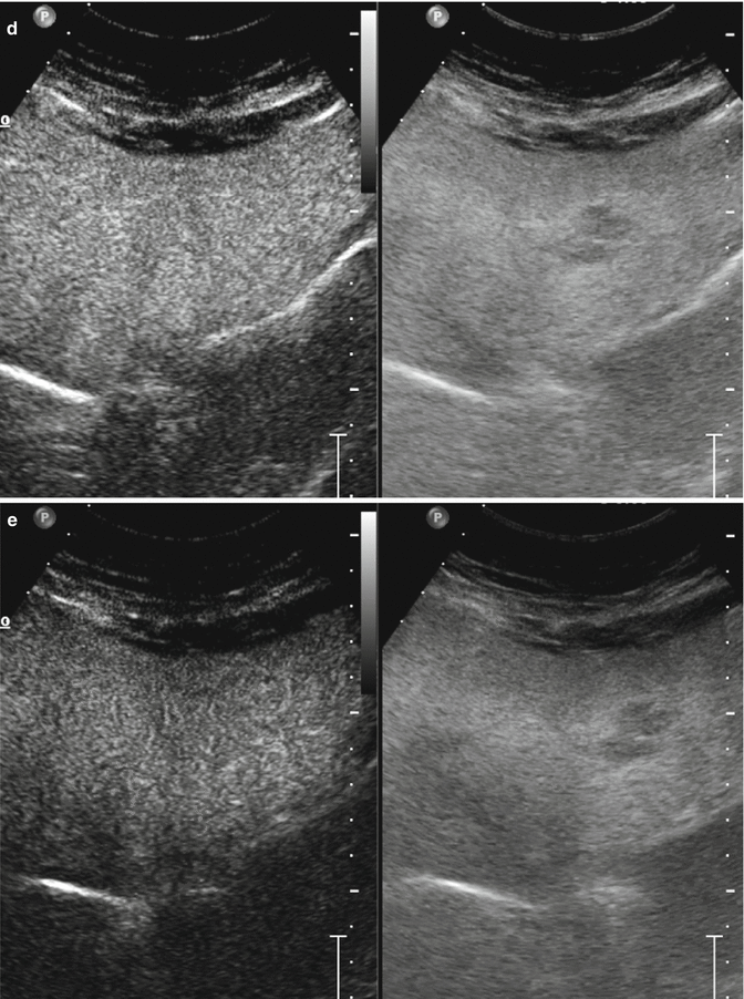
Focal fatty changes—presenting as hyperechoic areas in otherwise normal liver—and focal sparing areas—occurring as hypoechoic areas in a diffuse fatty “bright liver”—usually show no differences in contrast uptake in comparison with adjacent liver parenchyma. Consequently, at CEUS, these “pseudolesions” do not show contrast enhancement during the arterial phase and become isoechoic in comparison with the surrounding liver parenchyma in the extended portal-venous phase [13].
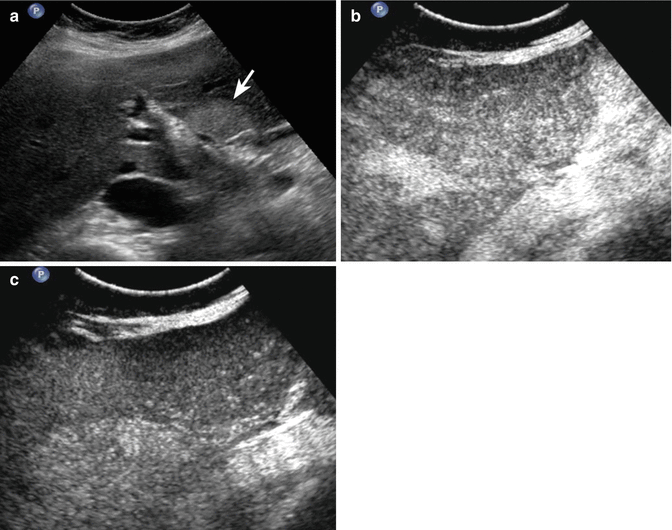

Fig. 4.3
Focal fatty area in a 58-year-old man. (a) At baseline US, an hyperechoic area sized 3.5 cm is detected in segment IV (arrow). (b, c) At CEUS, the area is evident neither in the arterial phase (b) nor in the late phase (c)

