Fig. 11.1
Endoscopic view of a superficial urothelial lesion of the left renal pelvis with a fiber optic (left) and a digital (right) ureteroscope
Moreover, an adequate staging of UUT-TCC patients can be hampered by technical difficulties in obtaining reliable pathological data with the ureteroscopic biopsy [3, 4]. Finally, not all the urothelial malignant lesions are always visible using white light, which represents the standard source of light of the standard endourological procedures. In the field of bladder cancer, enhanced imaging techniques are being developed with the aim of increase the diagnostic accuracy of cystocopy and improve the outocome of transurethral resection of the bladder (TURB) [5]. Nevertheless, these new technologies are being applied to the endoscopic management of malignancies of the upper urinary tract, with promising results.
In this chapter we sought to describe the different enhanced imaging techniques available on the market and the preliminary results of these devices when applied to the treatment of UUT-TCC.
11.1 Narrow Band Imaging (NBI)
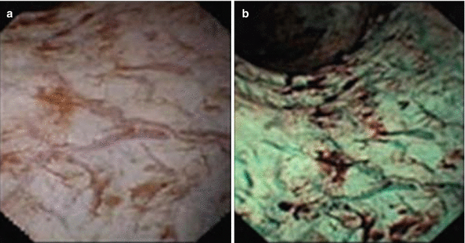
Narrow Band Imaging (NBI) system has been incorporated by Olympus in the endoscopic instrumentation. It consists of a new function for detecting suspicious lesions in the urinary tract which adopts the FWL system, an optical image technology that enhances the contrast between normal urothelium and cancer tissue by filtering white light into two bandwidths of 415 and 540 nm. These narrow bands of light are strongly absorbed by haemoglobin and penetrate the tissue surface only, increasing the visibility of mucosal vascular structures. As result, capillaries are displayed in brown and veins in the sub-surface are displayed in cyan on the operating monitor [6] (see Fig. 11.2).


Fig. 11.2
Normal urothelium. (a) Endoscopic view with white-light cystoscopy; (b) Narrow-band imaging cystoscopy
Due to the vascular nature of urothelial carcinoma, NBI enhances the contrast between superficial tumours and normal mucosa. Subjectively, NBI clearly shows the specific vascular architecture of UUT-TCC, creating the impression of a 3D visualisation of the malignant lesion and improving the definition of tumour edges.
Therefore, the spatial representation of tumours may affect the diagnostic accuracy of the endoscopic procedure, thus increasing the detection of all those malignant lesions not perfectly visible with white light.
This in turn may lead to improve the likelihood of complete tumour ablation, absence of tumour persistence at the end of the procedure and to reduce the risk of tumour recurrence. Data available in literature showed that cancer detection rate in patients undergoing NBI cystoscopy was higher than that of their white light cystoscopy counterparts [7]. Moreover, NBI cystoscopy performed after white light cystoscopy increased the detection of malignant lesions by 13–41 % [8, 9].
Finally, NBI has been proven to have a positive impact on the risk of tumour recurrence at 1 year after initial TURB. Indeed, it has been reported at 32.9 % in the NBI group versus 51.4 % in the white light group [10].
11.1.1 Incorporation in Video-Ureteroscopy
NBI system has been recently incorporated by Olympus in the digital ureteroscopes (namely, URF-V and URF-V2). As described in the field of bladder cancer, the current technique is expected to help clinicians in the diagnosis of early urothelial carcinoma and particular condition, such as carcinoma in situ. Indeed, due to the microvascular component characterising malignant lesion, flat lesion can be clearly distinguished from edematous lesions caused for instances by JJ stent, appearing as “frog’s eggs” (see Fig. 11.3).


Fig. 11.3
(a) Frog’s eggs; (b) Urothelial tumour with white-light ureterorenoscopy; (c) Urothelial tumour with narrow-band imaging system ‘frog’s eggs effect’; (d) Urothelial irritation due to JJ stent (white-light ureterorenoscopy); and (e) Urothelial irritation due to JJ stent (narrow-band imaging system) with no ‘frog’s eggs effect’
11.1.2 Preliminary Results of NBI Technology in the Diagnosis of UUT-TCC
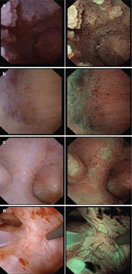
The first report regarding the use of such a technique in the field of endoscopic treatment of UUT-TCC came from Tenon University Hospital’s experience. Data coming from 27 patients were analyzed. Any area in the NBI mode which appeared as discordant by either blood vessel concentration or appearance (i.e., dotted, tortuous, large-calibre, abrupt-ending vessels) as compared to white-light mode was considered as an abnormal appearance suspected of being malignant lesion. NBI technology allowed to diagnose and clearly visualise UUT-TCC and to identify the extended tumour limits (see Fig. 11.4).


Fig. 11.4
Examples of endoscopic views with narrow band inaging (NBI) (left) and white-light (WL) (right). (a) Multiple lesion in pyelo-caliceal left kidney. (b) small lesion more visible with NBI versus WL. (c) Extent limits better identified with NBI versus WL. (d) No tumour visible in WL. Papillary tumour detected at NBI
When comparing the endoscopic results with bioptic findings, there were 35 pathologically confirmed transitional cell tumours detected. NBI diagnosed five tumours missed at WL (14.2 %) and clearly identified the edges of three tumours (8.5 %), improving tumour detection rate by 22.7 % [11].
However, no specimens were taken from the extended margins of the tumours that were only visible by NBI, therefore the authors were not able to confirm the hypothesis that such a technique helps clinicians to discriminate tumour limits.
Taken together, these preliminary findings may be the basis for developing further studies aimed at confirming the benefit of NBI not only in the diagnosis but also in the long term treatment of UUT-TCC. Indeed, the impact of NBI technology on the risk of tumour recurrence and progression over time in these patients has still to be determined.
11.2 Photodynamic Diagnosis (PDD)
Photodynamic diagnosis (PDD) relies on fluorescence produced by substances with particular properties called fluorochromes which can be localized into abnormal tissue. This technique, initially employed for the diagnosis of other malignancies, such as lung cancer, skin cancer, upper aerodigestive tract cancer, has been increasingly used in the urological field to identify urothelial malignant lesions. PDD is able to improve the diagnosis of all those tumours which would not be clearly seen with standard endoscopic procedure due to their flat shape and/or small size [12].
The fluorescence occurs when the outer electrons of the fluorochrome return at their own ground state after having been excited by the absorption of a photon of appropriate wavelength. This technique has been designed as a combination of a photosensitizing drug, delivered orally or topically and spontaneously absorbed by abnormal cells, and a specific instrumentation equipped with a source of light able to excite the substance itself by using specific wavelengths, thus enhancing specific fluorescence staining, and optical filters able to detect the fluorescent signal.
Among the substances available for PDD in the urological field, 5-aminolevulinic acid (5-ALA) is the most widely used.
11.2.1 5-ALA and Derivatives
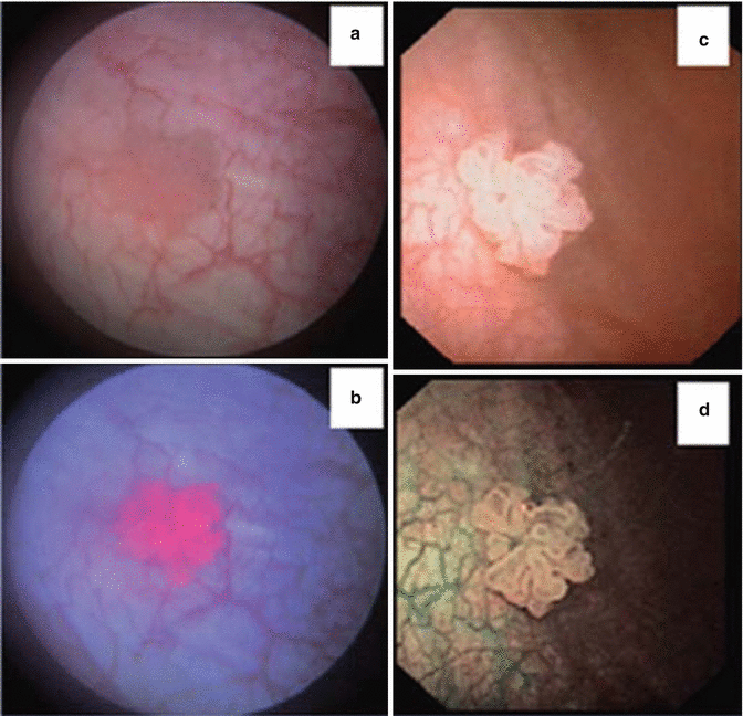
5-ALA plays a natural role in the biosynthetic pathway of heme. Indeed, it is the metabolic precursor of protoporphyrin IX (PpIX), which is the only fluorescent substance in the pathway. The step from protoporphyrin IX (PpIX) to heme, which include the insertion of a ferrous ion into the porphyrin ring, is the critical bottleneck of the whole pathway. Therefore, an exogenous administration of 5-ALA will induce elevated intracellular level of PpIX and consequently an increased fluorescence staining activity arising from neoplastic or highly proliferating cells [13, 14]. This phenomenon has been used by clinicians to better identify malignant and premalignant lesions during endourological procedures. The fluorescent-mode light used is the blue light, which serves as a reference for the normal urothelium, while the tissue accumulating the compound appears as red (see Fig. 11.5).


Fig. 11.5


Comparison of endoscopic visualization of a urothelial tumour with classic white-light (a), after 5-aminolevulanic acid instillation and blue light (b), with a white-light digital endoscope (c), and with NBI (d)
Stay updated, free articles. Join our Telegram channel

Full access? Get Clinical Tree


