Endoscopic ultrasound (EUS) has evolved as a useful technique for imaging and intervention in the colon and rectum. This article reviews the clinical applications of EUS for imaging and intervention in colorectal cancer, with an emphasis on the most recent clinical studies.
Endoscopic ultrasound (EUS) has evolved as a useful technique for imaging and intervention in the colon and rectum. Instruments, manufactured by various companies, for imaging the colorectal area include radial (cross-sectional) and linear (longitudinal) flexible endoscopic ultrasound echoendoscopes, rigid endorectal probes, and high-frequency catheter miniprobes that can be passed through the biopsy channel of conventional endoscopes. Interventional EUS procedures require use of the linear array echoendoscopes to permit visualization of a needle along its length. This article reviews the clinical applications of EUS for imaging and intervention in colorectal cancer, with an emphasis on the most recent clinical studies.
Artifacts in Colorectal Endoscopic Ultrasound
Artifacts in colorectal EUS include pseudomasses created by beam thickness during imaging of rectal folds and mirror image reflection at an intraluminal fluid level . Simulation of malignant infiltration may occur because of beam thickness, attenuation, or refraction. The understanding of the physical properties of ultrasound, with recognition of these artifacts, is important to avoid EUS misinterpretation.
Endoscopic Ultrasound in Rectal Cancer
EUS is a highly useful technique for preoperative local staging of rectal cancer to help determine the type of surgery required and whether preoperative neoadjuvant chemoradiation is needed. Savides and Master concisely summarized the indications for EUS in rectal cancer based on therapeutic impact, stratified according to cancer stage. Indications for EUS in rectal cancer include:
- •
To determine the suitability for endoscopic mucosal resection or transanal excision (if the lesion is T1 by EUS) for a large polyp or small rectal cancer
- •
To determine whether preoperative chemotherapy and radiation are necessary for a large, rectal cancer (T2: radical resection, T3-4 or N1: preoperative chemoradiation followed by radical resection)
- •
Surveillance after surgery for rectal cancer
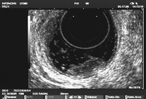
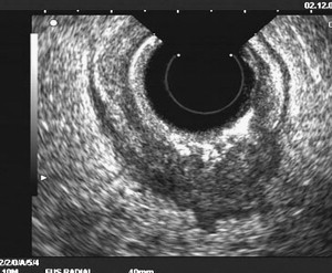
Glancy and colleagues analyzed the accuracy of EUS for selection for local excision by transanal endoscopic microsurgery (TEM) in 156 patients who had rectal neoplasia ( Fig. 1 ). EUS (uT stage) was compared with the postoperative histopathological stage of the resected specimens (pT stage). Of the 62 patients undergoing TEM, three were overstaged, and none were understaged by EUS (95% overall accuracy). Among the other 94 patients undergoing an alternative procedure, the accuracy of EUS at predicting advanced disease was 89%, and the overall accuracy was 92%. The accuracy of EUS for assessing local depth of invasion of rectal carcinoma (T stage) ranges from 80% to 95% ( Fig. 2 ) . This accuracy compares favorably with the accuracy of CT of 65% to 75% and the accuracy of MRI of 75% to 85% . Concerning T stage, the major limitation of EUS is overstaging of T2 tumors ; peritumorous inflammation cannot be distinguished from malignant tissue by ultrasound. On the other hand, the inability to detect microscopic malignant infiltration can lead to understaging. Stenotic rectal cancers sometimes are staged suboptimally by EUS because of an inability of the echoendoscope to traverse the stenosis , but stenotic tumors usually are advanced lesions.


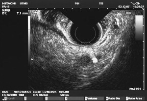
The accuracy of EUS for lymph node staging is less than that for T staging, because the echo features of benign/inflammatory and malignant lymph nodes overlap. It ranges from 70% to 75%. This accuracy compares favorably with that for CT (55% to 65%) and with that for MRI (60% to 65%) . Round, echo-poor or hypoechoic lymph nodes that are more than 5 mm in diameter are considered suspicious for metastases in rectal cancer ( Fig. 3 ) . EUS-guided fine needle aspiration (FNA) ( Fig. 4 ) can improve the accuracy of lymph node staging, but this can be done only for lymph nodes not immediately adjacent to the primary tumor, because traversing of the EUS FNA needle through the primary tumor will lead to spurious false-positive results.

Numerous studies have compared EUS with MRI and other imaging modalities. A recent study compared the ability of EUS versus either body coil MRI or phased-array coil MRI to locally stage rectal carcinoma before surgery in 49 patients . The EUS and MRI findings were compared with the histologic findings on the surgical specimen. For local T staging, the accuracy of EUS was 70%; the accuracy of body coil MRI was 43%, and the accuracy of phased-array coil MRI was 71%. For N stage, the accuracy of EUS, body coil MRI, and phased-array coil MRI was 63%, 64%, and 76%, respectively. For T staging, EUS had the best sensitivity (80%), but the same specificity (67%) as phased-array coil MRI. For N stage, phased-array coil MRI had the best sensitivity (63%), but the same specificity (80%) as the other methods. In this study, EUS and phased-array coil MRI provided similar results for assessing T stage. No method provided satisfactory assessments of local N stage, although phased-array coil MRI was marginally better in assessing this important parameter.
Endoscopic Ultrasound in Rectal Cancer
EUS is a highly useful technique for preoperative local staging of rectal cancer to help determine the type of surgery required and whether preoperative neoadjuvant chemoradiation is needed. Savides and Master concisely summarized the indications for EUS in rectal cancer based on therapeutic impact, stratified according to cancer stage. Indications for EUS in rectal cancer include:
- •
To determine the suitability for endoscopic mucosal resection or transanal excision (if the lesion is T1 by EUS) for a large polyp or small rectal cancer
- •
To determine whether preoperative chemotherapy and radiation are necessary for a large, rectal cancer (T2: radical resection, T3-4 or N1: preoperative chemoradiation followed by radical resection)
- •
Surveillance after surgery for rectal cancer
Glancy and colleagues analyzed the accuracy of EUS for selection for local excision by transanal endoscopic microsurgery (TEM) in 156 patients who had rectal neoplasia ( Fig. 1 ). EUS (uT stage) was compared with the postoperative histopathological stage of the resected specimens (pT stage). Of the 62 patients undergoing TEM, three were overstaged, and none were understaged by EUS (95% overall accuracy). Among the other 94 patients undergoing an alternative procedure, the accuracy of EUS at predicting advanced disease was 89%, and the overall accuracy was 92%. The accuracy of EUS for assessing local depth of invasion of rectal carcinoma (T stage) ranges from 80% to 95% ( Fig. 2 ) . This accuracy compares favorably with the accuracy of CT of 65% to 75% and the accuracy of MRI of 75% to 85% . Concerning T stage, the major limitation of EUS is overstaging of T2 tumors ; peritumorous inflammation cannot be distinguished from malignant tissue by ultrasound. On the other hand, the inability to detect microscopic malignant infiltration can lead to understaging. Stenotic rectal cancers sometimes are staged suboptimally by EUS because of an inability of the echoendoscope to traverse the stenosis , but stenotic tumors usually are advanced lesions.
The accuracy of EUS for lymph node staging is less than that for T staging, because the echo features of benign/inflammatory and malignant lymph nodes overlap. It ranges from 70% to 75%. This accuracy compares favorably with that for CT (55% to 65%) and with that for MRI (60% to 65%) . Round, echo-poor or hypoechoic lymph nodes that are more than 5 mm in diameter are considered suspicious for metastases in rectal cancer ( Fig. 3 ) . EUS-guided fine needle aspiration (FNA) ( Fig. 4 ) can improve the accuracy of lymph node staging, but this can be done only for lymph nodes not immediately adjacent to the primary tumor, because traversing of the EUS FNA needle through the primary tumor will lead to spurious false-positive results.
Numerous studies have compared EUS with MRI and other imaging modalities. A recent study compared the ability of EUS versus either body coil MRI or phased-array coil MRI to locally stage rectal carcinoma before surgery in 49 patients . The EUS and MRI findings were compared with the histologic findings on the surgical specimen. For local T staging, the accuracy of EUS was 70%; the accuracy of body coil MRI was 43%, and the accuracy of phased-array coil MRI was 71%. For N stage, the accuracy of EUS, body coil MRI, and phased-array coil MRI was 63%, 64%, and 76%, respectively. For T staging, EUS had the best sensitivity (80%), but the same specificity (67%) as phased-array coil MRI. For N stage, phased-array coil MRI had the best sensitivity (63%), but the same specificity (80%) as the other methods. In this study, EUS and phased-array coil MRI provided similar results for assessing T stage. No method provided satisfactory assessments of local N stage, although phased-array coil MRI was marginally better in assessing this important parameter.
Three Dimensional Endoscopic Ultrasound
Conventional two-dimensional (2D) EUS provides limited spatial information. Three-dimensional (3D) EUS image reconstruction may improve the accuracy of EUS and decrease errors in staging. Kim and colleagues studied both 3D and 2D EUS for staging rectal cancer in 33 patients. Accuracy of 3D EUS was 90.9% for pT2 and 84.8% for pT3, whereas that of conventional EUS was 84.8% and 75.8%, respectively. Lymph node metastasis was predicted accurately by 3D EUS in 28 patients (84.8%), but was predicted accurately in only 22 patients (66.7%) by 2D EUS.
Kim and colleagues recently reported a study comparing the efficacy of 3D EUS with that of 2D EUS and CT for staging of rectal cancer. Eighty-six rectal cancer patients were evaluated before surgery by 2D EUS, 3D EUS, and CT. EUS imaging was performed with rigid rectal probes. The accuracy of T staging was 78% for 3D EUS, 69% for 2D EUS, and 57% for CT, while the accuracy of detecting lymph node metastases was 65%, 56%, and 53%, respectively. Examiner errors were the most frequent cause of misinterpretation, occurring in 47% of 2D EUS examinations and 65% of 3D EUS examinations. By eliminating examiner errors, the accuracy rates of T staging and lymph node evaluation could be improved to 88% and 76%, respectively, for 2D EUS, and could be improved to 91% and 90%, respectively, for 3D EUS. Poorly differentiated or mucinous rectal tumors with adverse prognostic factors were closely associated with infiltration grade detected by 3D EUS in this study . The 3D reconstruction of rectal tumors revealed conical protrusions along the deep margins with the number of cones closely correlating with infiltration grade, advanced local T stage, and presence of lymph node metastases.
Giovannini and colleagues recently reported the staging of rectal cancer in 35 patients using a new software program for 3D EUS that can be used with electronic radial or linear rectal probes. No differences were evident with 3D EUS versus 2D EUS for superficial tumors (T1 and T2N0). In 6 of 15 patients classified as having T3N0 lesions, however, 3D EUS revealed malignant lymph nodes, a finding that was confirmed surgically in five of the six cases. Three-dimensional EUS also correctly assessed the degree of infiltration of the mesorectum, demonstrating complete invasion of the mesorectum in eight cases. These findings were confirmed by surgery in all the cases. Two-dimensional EUS was accurate for T and N staging in 25 of 35 rectal tumors (71.4%), whereas 3D EUS was accurate in 31 of 35 cases (88.6%). The authors concluded that 3D EUS provided more precise staging of lesions and better definition of the mesorectal margins in patients who had rectal cancer; this difference impacted on the therapeutic decisions.
Stay updated, free articles. Join our Telegram channel

Full access? Get Clinical Tree


