Endoscopic Evaluation of Lesions With Image-Enhancement Technologies
Narrow Band Imaging, i-Scan, Flexible Spectral Imaging Color Enhancement, and LASEREO
Shiaw-Hooi Ho, MD, MMED, AM
Introduction
Conventional chromoendoscopy (CE) with special dyes, such as indigo carmine, methylene blue, Lugol’s iodine, or crystal violet, provides an invaluable tool in the detection and assessment of luminal gastrointestinal (GI) lesions. Not only does it improve the detection rate of lesions through color and morphological enhancement, but it also allows histologic prediction, especially when used in conjunction with magnifying endoscopy. However, the main drawbacks include the cumbersome preparation and instillation of dyes, additional procedural time, and lack of availability of some of these agents in many centers.
Newer endoscopes are equipped to enhance imaging first by using a different illumination light source. This is achieved by either placing a filter to a broad-spectrum light source or using a light source that generates narrow-spectrum illumination. This concept of illumination with a narrow-spectrum wavelength is collectively called narrow-spectrum imaging (NSI), and it is the cornerstone of optical enhancement or optical CE. Second, through digital imaging, images can be enhanced by reducing “noise,” increasing contrast, sharpness, brightness, color, tone, etc; this is commonly referred to as digital enhancement or digital CE. Optical digital CE or equipment-based image-enhanced endoscopy (e-IEE) has the advantage of being the more convenient-to-use image-enhancement technologies compared to conventional dye-based CE. Utilizing filtered illumination light and/or digital manipulation, e-IEE permits detailed visualization of the microsurface and microvessel structures, which ultimately aids in the endoscopic detection and characterization of luminal GI lesions.
Such a concept of enhancement of vascular visualization using filtered illumination light was introduced in other fields of medicine almost a century ago. In 1913, Vogt described the use of a green (red-free) filter to examine the retinal vasculature, which is still practiced even in modern-day ophthalmology. In fact, the use of filtered illumination light in GI endoscopy came rather late. It was not until 5 decades after the invention of the flexible gastroscope that filtered illumination light was finally introduced by Gono and colleagues.1 And with it came the revolution in the field of diagnostic imaging in GI endoscopy. The details of each of these technologies are described in the following sections.
Technical Aspects
Light Source and Image Sensor
Conventional lamps, such as incandescent and fluorescent lamps, are common in daily household use. The incandescent lamp employs a tungsten filament that heats up and glows when electric current flows through it. A fluorescent lamp, on the other hand, generates short-wavelength ultraviolet light when an electric current excites the mercury vapor contained in the fluorescent tube. This generated ultraviolet light then causes the phosphor coating on the inner side of the lamp to glow and generate white-light illumination. Phosphor is a chemical compound that glows upon excitation by various electromagnetic waves. The best known phosphor materials are copper-activated zinc sulfide and silver-activated zinc sulfide. The commonly used light source in endoscopic systems is the xenon lamp. This lamp is essentially a gas discharge lamp made up of tungsten electrodes contained within a tube filled with highly pressurized ionized xenon gas. When an electric field is applied, movement of electrons causes the electrons in the outer orbit of the xenon atoms to be detached transiently. The return of the detached electrons to their lower-energy native orbits (resting state) releases energy in the form of light (which closely resembles that of natural sunlight). Other examples of newer light sources employed in endoscopic systems are light-emitting diodes (LEDs) and light amplification by stimulated emission of radiation (LASER). An LED is made up of P-type and N-type semiconductors combined to form a diode. When an electric field is applied, movement of electrons from N-type to P-type semiconductors across the junction causes the electron to lose energy in the form of light to fit onto the lower energy orbit of the P-type semiconductor. The light generation by LED is more efficient, as less energy is wasted on heat generation. Laser technology involves a similar concept of light emission following the return of excited electrons to their resting state but the generated light undergoes further amplification as it get reflected by the 2 mirrors within the laser tube. Light generated both by LED and laser is monochromatic, but laser is unidirectional and coherent while LED is not. As for the image sensors, there are 2 types of commonly available image sensors, namely the charged-coupled device (CCD) and the complementary metal oxide semiconductor (CMOS). CCD sensors generally produce higher-quality images with lower image noise while CMOS sensors consume less power and have a faster processing speed. Conventional endoscopes employ CCD sensors while newer endoscopes utilize CMOS sensors as the quality of CMOS sensors has improved tremendously over the years. The pixel is the smallest unit in each image sensor or display device. An image sensor or display device is considered to be high resolution or high definition when it delivers at least 720 horizontal lines of pixel information in a progressive scanning manner. In a high-resolution (HR) image sensor with a 16:9 aspect ratio, there will be at least 1280 vertical lines and 720 horizontal lines of pixels, giving rise to a total of at least 921,600 pixels.
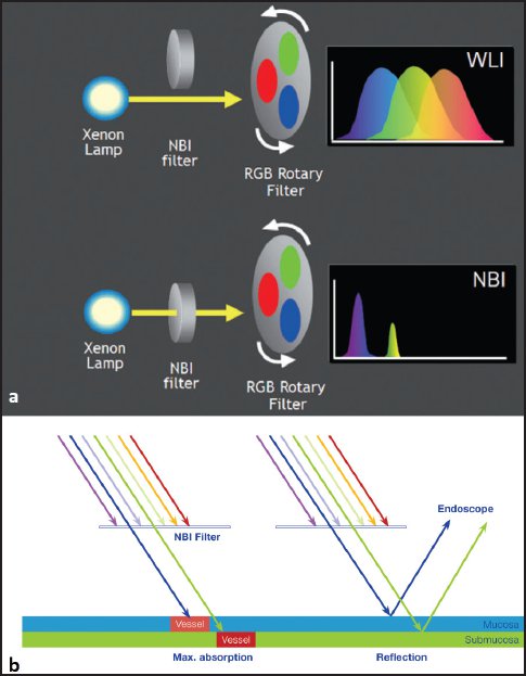
Figure 3-1. (A) An NBI filter placed in front of the light source limits the light spectrum and generates NSI. (B) Light at 415 nm wavelength (blue) is absorbed by the most superficial microvasculature while light at 540 nm wavelength (green) is absorbed by deeper vasculature such as the arteriole and venule. WLI, white-light imaging. (Reprinted with permission from Kazuhiro Gono, PhD and Olympus.)
Narrow Band Imaging
Narrow Band Imaging (NBI) was officially launched by Olympus in 2005 and was the first introduced e-IEE modality (optical digital CE).1 A light spectrum generated by a xenon lamp is filtered by mechanical filters so the output wavelengths fall in the range of 415 nm and 540 nm (Figure 3-1A). Light in the 415 nm wavelength is absorbed by red blood cells (hemoglobin specifically) traveling in the most superficial microvasculature (eg, intrapapillary capillary loop in the esophagus, subepithelial capillary network [SECN] in the stomach, and meshed capillary vessels [MCVs] in the colon), resulting in the dark appearance of superficial blood vessels, while light in the longer 540 nm wavelength is absorbed by the deeper vasculature, such as arterioles and collecting venules (Figure 3-1B). This effect leads to an increase in the contrast and “visibility” of the superficial microvasculature structures. In its truest sense, NBI is an optical CE technology.
Now in its second generation, the discharge lamp has been upgraded to a brighter type, the sensitivity of the image sensor (higher sensitivity means better ability to capture an image in darker conditions) has been improved, and the image processing noise (generally, more image noise is generated when an image is taken in darker ambience) has been further reduced. All of these improvements result in brighter and better NBI images.

Figure 3-2. In the Fujifilm LASEREO system, 2 laser beams are used to provide illumination. One blue laser beam provides the narrowed spectrum at 410 nm (BLI) and another laser beam at 450 nm is used to excite a phosphor, which gives rise to white-light illumination. (Reprinted with permission from the Endoscopy Systems Division of Fujifilm.)
Flexible Spectral Imaging Color Enhancement and i-Scan
Flexible Spectral Imaging Color Enhancement (FICE) and i-Scan are the first-generation e-IEEs introduced by Fujifilm (2005) and Pentax (2007), respectively. Image captured by white-light imaging (WLI) is digitally enhanced using proprietary software. Generally, these enhancements include adjustment of brightness, contrast, sharpness, color, tone, etc. Hence, with these postimaging processing technologies, there is no actual change in the illumination light. These technologies are referred to as digital CE as opposed to optical CE of NBI.
FICE, known earlier as Fujinon Intelligent Chromo-Endoscopy, permits the advanced calculation of each WLI image and determines the value of each wavelength of light in each individual pixel in the image sensor. Thus, FICE is able to put out a single-wavelength image with wavelength determined by the operator in increments of 5 nm from a range of 400 to 700 nm. Furthermore, sharpness and tonal adjustment are available for a better viewing experience.
Similarly, i-Scan provides an enhanced view of the mucosal surface and vasculature via such a modification on its WLI image. Three enhancement modes are available: surface, contrast, and tone enhancements (cyan and green). Based on the selection of these enhancement modes, 3 standard default presettings are available: i-Scan 1, 2, and 3.
Blue Laser Imaging and Blue Light Imaging
Realizing the shortcoming of digital CE as compared with optical CE, Blue Laser Imaging (BLI) was introduced by Fujifilm in 2012 (Figure 3-2). It is the first endoscopic system (LASEREO, Fujifilm) that utilizes 2 laser beams of 410 nm and 450 nm wavelengths as the source of light.2 The 410 nm wavelength is readily absorbed by hemoglobin in the most superficial microvasculature similar to NBI of 415 nm. The 450 nm wavelength serves to excite a phosphor, which in turn generates white light. Illumination from BLI and WLI can be achieved by changing and switching the intensity of these 2 laser beams. A BLI system utilizing LED as a light source was recently introduced (Figure 3-3A). In this new system (Eluxeo 7000, Fujifilm), blue-violet, blue, green, and red LEDs provide illumination. The combination of the illumination from these 4 LEDs generates WLI. The BLI mode in this new system is simply provided by the illumination from the blue-violet LED, which generates light in the 410 nm wavelength territory. BLI bright mode is BLI mode with brighter illumination. This was made available as an add-on to the original BLI mode to allow visualization of distant structures, especially when BLI is intended to be used as a detection tool.

Figure 3-3. (A) In the Fujifilm Eluxeo 7000 system, 4 LEDs (blue-violet, blue, green, and red) are used to generate white-light illumination. (B) In narrow-spectrum mode (BLI), the illumination at 410 nm is provided by the blue-violet LED. In linked color imaging, the illumination from the region of blue-violet is enhanced while the illuminations from the blue, green, and red regions are similar to WLI, giving rise to the enhanced microsurface and microvasculature appearance in the familiar white-light viewing environment. (Reprinted with permission from the Endoscopy Systems Division of Fujifilm.)
Related posts:
 Introduction and History of Endoscopic Resection Techniques
Endoscopic Resection Outcomes and Postresection Surveillance of Barrett-Associated Neoplasia
Introduction and History of Endoscopic Resection Techniques
Endoscopic Resection Outcomes and Postresection Surveillance of Barrett-Associated Neoplasia
 Electrosurgical Equipment and Principles of Electrosurgery
 Endoscopic Resection Outcomes and Postresection Surveillance in the Colon
 Prevention and Management of Postendoscopic Resection Luminal Strictures
 Endoscopic Submucosal Dissection Technique to Conquer Difficult Cases
Stay updated, free articles. Join our Telegram channel

Full access? Get Clinical Tree


