Fig. 8.1
Choline PET/CT images of a 69-year-old patient with biochemical recurrence (PSA serum level 2.3 ng/ml) after radical prostatectomy for prostate cancer. (a) Axial coronal fused PET/CT image showing an uptake of the radiotracer at the lateral arch of the III right rib and at the level of left para-aortic and homolateral external iliac lymph nodes. No uptake at the level of the post-prostatectomy bed was found. (b) Sagittal fused PET/CT image showing a focal uptake at occipital bone. (c) Axial fused PET/CT image displaying the presence of two focal uptake of radiotracer at the soma of T6 and at the lateral arch of the III right rib. All these findings are consistent with bone and lymph nodes metastases
The accuracy of choline PET/CT in staging and restaging of PC has been assessed by several studies [16].
Choline is a component of phosphatidylcholine, an important element of cell membranes; as known, biosynthesis of the cell membrane is very fast in tumour tissues and the up-regulation of choline kinase activity, particularly increased in PC cells, induces a higher uptake of choline [17].
This radiopharmaceutical has been employed in detecting various tumours, such as brain lesions [18], lung carcinoma [19] and bladder cancer [20].
However, its main clinical use is the study of PC, as demonstrated by numerous studies published in recent years. The [11C]-choline, in fact, is taken up in the pelvis exclusively by prostatic tissue and this property is retained by the neoplastic tissue. The [11C]-choline has also a negligible elimination through the urinary tract and the prostate is the only organ to have a significant uptake of the tracer in the pelvic region.
Regarding 18F-FDG, some studies have been showed that PET/CT with choline can detect more metastatic lymph nodes and bony metastatic lesions than 18F-FDG PET/CT in PC patients [21].
Furthermore, Picchio et al. [22] in a study showed that choline PET/CT identified more lesions suspected for neoplastic relapse (42 %) compared to 18F-FDG PET/CT (27 %) and demonstrated that choline PET is more accurate in identifying both local and distant recurrence.
There are several studies on the role of choline PET in the detection of primary tumour in the prostate gland [23, 24] and its use in the staging of prostatic disease before treatment [25, 26], especially in relation to PSA value [27]. However, its role in this field is still not clear, since the uptake of choline can occur in some benignant conditions, such as prostatic hyperplasia or prostatitis.
Therefore, the restaging of PC can be the main field of application of this imaging modality. In particular, the main role of choline PET/CT is represented by its ability to identify the location of the recurrence of the disease in the patients who had already undergone radical surgical treatment for prostate cancer and that present a biochemical relapse [28, 29].
Heinisch et al., in a single-centre retrospective study [30], analysed 31 patients after RP and found that 8/17 patients (47 %) with biochemical recurrence and with PSA <5 ng/ml, had positive results at 18F-choline PET/CT. Furthermore, in this paper, they found malignancy in 7/8 men confirmed by either biopsy or the course of the disease.
Rinnab et al. [31], in a single-centre retrospective study, analysed 50 patients with biochemical recurrence after primary therapy for PC (mean PSA serum levels: 3.62 ng/ml; range 0.5–13.1 ng/ml); the authors considered the sensitivity and specificity of 11C-choline PET/CT only in patients with PSA < 2.5 ng/ml, reporting 91 % and 50 %, respectively. In another single-centre retrospective study, Rinnab et al. [32] enrolled 41 patients with biochemical failure following RP (mean PSA was 2.8 ng/ml; range 0.41–11.6 ng/ml); overall, the sensitivity of 11C-choline PET/CT was 93 %, specificity 36 %, PPV 80 % and NPV 67 %.
Castellucci et al. [33] enrolled 190 patients with biochemical recurrence after RP (mean PSA serum level 4.2 ng/ml; range 0.2–25.4 ng/ml) and found an overall sensitivity of 11C-choline PET/CT of 73 % and specificity of 69 %. The same group [34], in another single-centre retrospective study, analysed 102 patients with biochemical relapse after RP (PSA serum level ranging from 0.2 to 1.5 ng/ml) submitted to 11C-choline PET-CT examination; all suspected local recurrences at PET/CT have been confirmed at TRUS-guided biopsy. Regarding local recurrence, the sensitivity of 11C-choline PET-CT was 53.8 % and specificity was 100 % (no false positive were recorded). Giovacchini et al. [35] in a single-centre retrospective study analysed 170 patients with biochemical failure after RP submitted to 11C-choline PET/CT; the mean PSA serum level at the time of the exam was 3.24 ng/ml (range 0.23–48.6 ng/ml) and mean PSAdt was 9.37 months. PET-CT showed a sensitivity of 87 %, specificity of 89 %, PPV of 87 %, NPV of 89 % and accuracy of 88 %. In this study, PET-CT positive findings were confirmed using histological analysis of lymph node specimen, anastomosis biopsy of the urethra/bladder neck, progression on PET-CT follow-up studies associated with increased PSA level, confirmation with conventional imaging, disappearance or sizable reduction of choline uptake after local or systemic treatment and PSA decrease greater than 50 % after selective irradiation of the unique site of pathological choline uptake.
In a second single-centre retrospective study, Giovacchini et al. [36] enrolled 358 patients with biochemical recurrence after RP: the mean PSA value was 3.77 ng/ml (range 0.23–45.2 ng/ml). 11C-choline PET-CT was performed in all patients and results were validated by histological analysis. Authors reported an overall sensitivity of 85 %, specificity 93 %, PPV 91 %, NPV 87 % and accuracy 89 %.
In the third single-centre retrospective study, Giovacchini et al. [37] from a database of 2,124 patients, retrospectively analysed 109 patients with biochemical recurrence (mean PSA before imaging of 1.31 ng/ml, range 0.22–16.76 ng/ml) who underwent 11C-choline PET/-CT. They reported positive findings at 11C-choline PET-CT in 12/109 patients, as local recurrence in 4 patients and as pelvic nodal metastases in 8 cases.
A relevant limit of all these studies is the lack of information on local recurrence dimensions.
Reske et al. [38], in a single-centre retrospective study, analysed 49 patients who underwent 11C-choline PET-CT examination with mean PSA level 2 ng/ml and median maximal diameter of the lesions of 1.7 cm (range 0.9–3.7 cm); TRUS biopsy was used to validate the results. They concluded that 11C-choline PET-CT had a sensitivity of 73 %, specificity 88 %, PPV 92 %, NPV 61 % and an accuracy of 78 %.
Up to now, the overall choline PET/CT sensitivity in detecting sites of PC relapse ranges between 38 % and 98 %. It has been demonstrated that choline PET/CT technology positive detection rate improves with increasing PSA values.
The most important feature provided by all the cited studies on this topic is linked to the strict relationship between choline PET/CT detection rate and PSA value in restaging PC patients. In the last decade, various authors have been proposed some cut-off values to help in discriminate those patients who can potentially benefit by a choline PET/CT scan; Cimitan et al. [39] proposed that a PSA cut-off value higher than 4 ng/ml is more probably associated to the possibility to detect distant metastases.
It has been found more higher is the value of PSA at the time of the scan, greater is the detection rate of choline PET/CT: 36 % for values of PSA <1 ng/ml; 43 % for PSA values between 1 and 2 ng/ml; 62 % for PSA values of between 2 and 3 ng/ml and 73 % if the PSA ≥3 ng/ml [40].
More recently, several authors proposed lower PSA cut-off values to individuate the patients to undergone choline PET/CT. Rinnab et al. proposed a cut-off value of 1.5 ng/ml but, generally, various authors are in agreement regarding a better sensitivity of the exam when performed in patients with PSA serum level higher or equal to 2 ng/ml [31, 32, 41].
More recently, the attention of the researchers has been focused on the potential role of PSA kinetics such as PSAdt, already cited, and PSA velocity (PSAve), a PSA derivative determined as linear regression of the PSA values over time [9].
In particular, standing to literature data, PSAdt and PSAve values are correlated with specific mortality risk of PC [42]. Additionally, it has been well reported that the risk of distant metastases in patients with biochemical failure after RP depends on PSA and PSAdt values. In particular, it has been showed that when PSAdt is more than 6 months, the risk of metastasis is less than 3 %, even with absolute PSA values of >30 ng/ml. If PSAdt is less than 6 months and PSA is >10 ng/ml, the risk of metastasis is near to 50 % [43].
Partin et al. [44] evaluated the usefulness of PSAve in predicting recurrence after RP and found that combining data relative to PSAve, Gleason score and pathological staging is helpful in distinguishing local recurrence from distant metastases.
Generally, a PSAdt the sensitivity of choline PET/CT is significantly higher in patients with a PSAve >2 ng/ml/year or a PSAdt ≤6 months [45]. In the cited study, in particular, the proposed PSAve cut-off of 2 ng/ml/year seemed to more accurately separate patients with a positive PET/CT scan from those with a negative scan, even if other authors suggest that the patients with a PSAve >1 ng/ml/year should be submitted to choline PET/CT [46].
In all cited studies, the very good detection rate or the sensitivity of choline PET/CT is often associated with distant metastases (for both lymph node and bone metastases) while the data about the diagnosis of local relapse are still discordant. In particular, when the mean PSA level is lower than <1.5 ng/ml, the detection rate of choline PET/CT for local recurrence is definitely poor, probably because of low PET spatial resolution limits (5–6 mm) which do not allow the detection of small lesions.
In a review article, Picchio et al. [41] confirm that the routine use of choline PET/CT for localisation of local relapse of PC cannot be recommended for PSA values <1 ng/ml.
In synthesis, standing to literature data, the choline PET/CT could play a role in management of PC patients, in particular during the restaging, with a good sensitivity regarding distant metastases and good detection rate in relationship to PSA value higher than 2 ng/ml, PSAdt lower than 6 months and PSAve higher than 2 ng/ml/year. To date, its role in detecting local recurrence in prostatic fossa after radical surgical treatment still remains unclear.
8.3 Mp-MRI
In the last 20 years, several progresses have been done in the use of MRI. High-field strength endorectal coil MRI is able to produce a morphological imaging of the prostate, particularly with T2-weighted imaging. Other recent complementary functional techniques that are Dynamic contrast-enhanced MRI (DCE-MRI), 1H-spectroscopic imaging (1H-MRSI) and Diffusion-weighted imaging (DWI) improve the staging and the detection of PC. DCE-MRI is a technique based on Gradient-Echo T1-weighted sequences used during the passage of a gadolinium contrast agent in order to assess the neoangiogenesis; therefore, it can detect those tumours in which an angiogenic pathway has been turned on [47]. DWI yields qualitative and quantitative information about tissue cellularity and cell membrane integrity. Intraductal and extracellular water molecules move freely. In PC, extracellular space is decreased; therefore, the water molecule movement is restricted and the so-called apparent diffusion coefficient values are low. DWI can be produced without the administration of exogenous contrast medium and can be considered the most practical and simple to use [48]. MRSI is a three-dimensional data set of the prostate, with volume elements (voxels) ranging from 0.24 to 0.34 cm [49]. This technique produces the relative concentration of metabolites within voxels, such as citrate, choline and creatine. Recent studies demonstrate that in PC citrate levels are reduced, creatine and particularly choline are elevated. The peak integral ratio of choline plus creatine to citrate can distinguish from PC tissue to healthy tissue [50]. On the basis of the literature, each voxel can be categorised as follows: fibrotic or scar tissue when the ratio is <0.2, residual healthy prostatic glandular tissue when ratio is >0.2 and <0.5, probably recurrent PC when ratio is >0.5 and <1 and definitively recurrent PC tissue when ratio is >1 [51]. MRSI technique is more complex when compared with DWI or DCE-MRI and it also requires longer acquisition times. Mp-MRI has the advantage to have a very good spatial resolution so as to localise and characterise PC, to detect very small lesions and to better differentiate from healthy to neoplastic areas. It is a complex technique and needs experienced and trained radiologists, in particular if MRSI is considered.
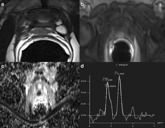
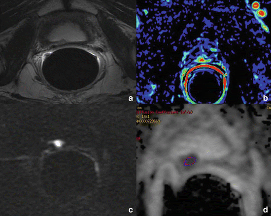
Recently, mp-MRI more than other imaging procedures (Figs. 8.2 and 8.3) has been proposed as a useful tool in the diagnosing of local recurrence of PC after RP [52].


Fig. 8.2
Multiparametric-MR images of a 64-year-old man with prostate-specific antigen progression (PSA serum level 0.75 ng/ml) after radical retropubic prostatectomy, with suspected local recurrence. (a) Axial T2-weighted fast spin-echo image shows a solid tissue of 1 cm in size on posterior perianastomotic location in front of the rectal wall at about 40 mm from the ureteral meatus which is slightly hyperintense compared to pelvic muscles. (b) Axial Gradient-echo T1-weighted image showing a remarkable enhancement of the pathological tissue. (c) Axial ADC map reconstructed from images obtained at b values of 0, 500 and 1,000 s/mm shows a dark area corresponding to the abnormal hyperintense tissue seen on T2-weighted images. (d) 1H-magnetic resonance spectroscopic imaging reveals a high choline peak with a choline-plus-creatine-to-citrate ratio greater than 0.9. All these findings are consistent with local recurrence

Fig. 8.3
Multiparametric-MR images of a 74-year-old man with prostate-specific antigen progression (PSA serum level 0.43 ng/ml) after radical retropubic prostatectomy, with suspected local recurrence. (a) Axial T2-weighted fast spin-echo image shows a solid nodular tissue of about 7 mm in size on the right posterior perianastomotic location in front of the rectal wall at about 12 mm from the ureteral meatus which is slightly hyperintense compared to pelvic muscles. (b) Axial Gradient-echo T1-weighted colour map image showing a remarkable enhancement of the pathological tissue. (c) Axial DWI image at b value = 1,000 s/mm showing marked restricted diffusion phenomena of water molecules. (d) Axial ADC map reconstructed from images obtained at b values of 0, 500 and 1,000 shows a dark area corresponding to the abnormal hyperintense tissue seen on T2-weighted images and hypervascular nodule seen on colour map. All these findings are consistent with locoregional relapse
Once biochemical progression of PSA serum level has been diagnosed after RP, it is essential for treatment planning to determine whether the recurrence has developed at local or distant sites, but the possibility of residual glandular tissue should be taken into account. In this setting, diagnostic imaging techniques are useful to differentiate local cancer recurrence from systemic relapse and to direct patients to the best therapeutic approach, that is, radiotherapy for local recurrence and hormone therapy for systemic disease [12]. Moreover, it is very important for radiation oncologists to differentiate residual prostatic healthy tissue from local recurrence because the dose of radiation therapy is different [53].
Choline PET/CT is recommended when PSA serum value is higher than 1 ng/ml because this technique has good sensitivity and specificity in detecting lymph nodes, distant recurrence and local recurrences after RP only in patients with high PSA values. Moreover, the ability of choline PET/CT in detecting local recurrence depends on lesion size, being usually higher if the lesion is more than 1 cm in diameter [54].
Although Ch-PET/CT is recommended for high PSA values, in patients with low biochemical alterations after RP (0.2 < PSA < 1 ng/ml) it is very important for radiation oncologists to exclude the presence of locoregional relapse.
Within mp-MRI, DCE-MRI has proved to be the most reliable technique in depicting locoregional relapse [55–58].
Up to now, there are several studies which demonstrate the usefulness of MRI in detecting local cancer recurrence. At present, mp-MRI after RP is indicated to diagnose small local recurrence and—thanks to functional imaging—to distinguish between residual glandular tissue and or/fibrosis and nodule recurrence; it may also be able to determine the aggressiveness of nodule recurrence. Panebianco et al. [59] compared ADC values of local recurrences with the histological results. The mean and standard deviation of ADC values were 0.5 ± 0.23 mm2/s for high-grade aggressiveness recurrence, 0.8 ± 0.09 mm2/s for intermediate-grade aggressiveness recurrence, 1.1 ± 1.17 mm2/s for low-grade aggressiveness relapse and the patients with a histological finding of residual glandular tissue had ADC values higher than 1.3 mm2/s (mean ADC values 1.4; range 1.3–1.7).
The perianastomotic fibrosis appears hypointense on T2-weighted images, with absent enhancement on DCE-MRI. After DCE-MRI, all benign nodules show signal enhancement of less than 50 % in the early phase, whereas all recurrences showed fast signal enhancement in the early phase followed by a plateau or washout. Recurrences appear as masses with intermediate to high signal intensity on T2-weighted images compared to pelvic muscles, enhancing after intravenous injection of contrast medium.
Stay updated, free articles. Join our Telegram channel

Full access? Get Clinical Tree


