Fig. 14.1
Treatment of a ureteral stone with distal ureteral stricture
Pre-operative Considerations
Patients with a history of difficult or impacted stones, multiple prior ureteroscopies, abdominal surgeries or radiation therapy are prone to ureteral stricture formation. A detailed history of prior stone events and surgical intervention should be obtained, including prior surgical complications. Pre-operative imaging with a non-contrast computed tomography (CT) scan may not suggest presence of a ureteral stricture in the setting of a ureteral stone. Urinary tract infections should be treated with antibiotics prior to any surgical intervention. It is also important to discuss with the patient realistic expectations of the surgery with the possibility of multiple procedures when the presence of a ureteral stricture and/or impacted stone is suspected.
Retrograde vs Antegrade Ureteral Access
In the setting of a concomitant ureteral stricture and stone, it may be difficult to gain retrograde access. Often times the stone is impacted at the location of the ureteral stricture making access even more challenging. An attempt should be made to pass a guidewire, such as the Sensor (Boston Scientific, Natick, MA, USA), under fluoroscopic visualization. If there is difficulty bypassing the stone a 5F ureteral access catheter should be passed under fluoroscopic guidance over the guidewire to a position just distal to the obstruction. The wire should be removed in order to perform a retrograde pyelogram (RPG) to delineate the anatomy. The ureteral access catheter will also straighten out the ureter distal to the obstruction allowing for the guidewire to project in a more favorable orientation. Using the imaging gained from the RPG a hydrophilic guidewire, such as a glidewire, should then be re-advanced through the ureteral access catheter to negotiate past the obstructing segment. It is important to carefully manipulate the guidewire with care in order to avoid ureteral perforation. Glidewires are often able to bypass an impacted stone or obstructing stricture and are less likely to cause a false passage or ureteral perforation. There are both straight and angled glidewires available. Once the glidewire is coiled within the renal pelvis, the ureteral access catheter should be passed over the wire and above the area of obstruction. If the urine draining from the access catheter appears to be infected then the procedure should be abandoned, a stent should be placed and treatment should be performed after appropriate antibiotics have eradicated the infection. Otherwise, the glidewire should then be exchanged for a stiffer guidewire to avoid the glidewire from inadvertently being removed.
If the above maneuver is not successful then the next step is placement of the guidewire under direct visualization. A guidewire should be left in place just below the area of obstruction and the ureteroscope, either semi-rigid if within the distal ureter or flexible if within the mid/proximal ureter, should be passed to the level of the obstruction. Under direct visualization the diameter of the stricture, size of the stone, and presence of stone impaction may be assessed. The guidewire may then be directed around the stricture and stone at a favorable appearing location. If stone impaction prevents this, then laser lithotripsy may be carefully performed until a guidewire is able to be passed. It is not recommended to attempt basketing the stone without a safety wire in place. In cases of failed retrograde access a percutaneous nephrostomy tube should be placed with plans for antegrade ureteroscopy.
Percutaneous antegrade treatment of ureteral stones is a secondary treatment after failed retrograde access to the stone. A stone positioned in the ureter just proximal to a ureteral stricture may be best approached in this manner before a standard endoureterotomy is performed. An upper-posterior or mid-posterior caliceal approach to percutaneous access offers the most direct route to the ureter. Flexible antegrade URS with laser lithotripsy may then be performed. A nephrostomy tube should be replaced to perform an antegrade nephrostogram post-operatively to evaluate the location and extent of the ureteral stricture for definitive surgical treatment.
After successful retrograde access the next step is determined by the location of the stricture that is compromising access to the stone. Due to the fragility of the proximal ureter, it is best to stent and allow for passive dilation rather than active dilation with a ureteral balloon dilator. A second-look URS may be done in 1–2 weeks, typically with an easily accessible ureter. Passive dilation is a safe and effective way to dilate the ureter; however, it does require two separate procedures. For distal ureteral strictures, a safety wire should be placed prior to balloon dilation. The safety wire may be placed with a dual lumen catheter, 8/10 dilator, ureteral access sheath, or under direct visualization with the semi-rigid ureteroscope.
Ureteral Balloon Dilation
Ureteral balloon dilators are recommended for dilation of ureteral strictures that compromise access to the ureteral stone. Use of balloon dilators are associated with increased post-operative pain and should only be used when necessary [1]. Balloon dilators range in length from 4 to 10 cm with various diameters, 12–15F, see Table 14.1 for specifications. The balloon has radiopaque markers at the proximal and distal ends, which are used to position the balloon over the working guidewire using fluoroscopy (Fig. 14.2). They should never be inflated over an obstructing ureteral stone due to the risk of ureteral perforation. Once the balloon is in proper position, it is inflated with half-strength contrast using a pressure gauge syringe. The balloons typically can withstand inflation pressures up to 17–20 atm; however dilation of the ureter typically can be achieved at lower pressures (4–6 atm). The lowest pressure necessary to dilate the ureter should be used to avoid ureteral perforation, and therefore it is important to inflate the balloon under fluoroscopic guidance. Typically a “waist” is seen in the balloon at the location of the stricture, and one should continue to inflate slowly until the “waist” disappears. The balloon is then deflated and removed, leaving the guidewire in place. Ureteroscopy may then be performed to treat the stone. Complications that can occur with balloon dilation include: balloon rupture from over inflation, mucosal injury, bleeding, and ureteral perforation if inflation is performed too rapidly or if higher pressures are used than necessary.
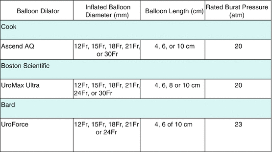
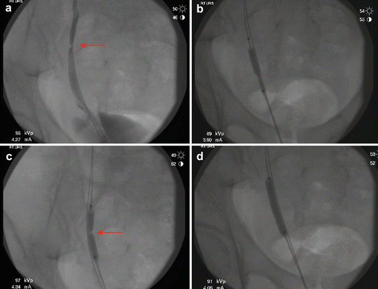
Table 14.1
Specifications of current ureteral balloon dilators
Fig. 14.2
 Radiation Exposure to the Patient and the Urologist
Shock Wave Lithotripsy for the Treatment of Ureteral Stones
Selecting the Appropriate Treatment Modality for Ureteral Calculi
Complications in the Treatment of Ureteral Stones: Prevention and Management
Radiation Exposure to the Patient and the Urologist
Shock Wave Lithotripsy for the Treatment of Ureteral Stones
Selecting the Appropriate Treatment Modality for Ureteral Calculi
Complications in the Treatment of Ureteral Stones: Prevention and Management
 Semirigid Ureteroscopy for Ureteral Calculi
 Tips and Tricks in the Treatment of Ureteral Stones


Balloon dilation of a ureteral stricture. (a) A retrograde pyelogram (RPG) is performed to evaluate the area of obstruction. The arrow indicates the location of the ureteral stricture. (b) The balloon has radiopaque markers at the proximal and distal ends, which is used to position the balloon over the working guidewire using fluoroscopy. Once the balloon is in proper position, it is inflated with half-strength contrast using a pressure gauge syringe. (c) It is important to inflate the balloon under fluoroscopic guidance to dilate the stricture with the lowest pressure required. Typically a “waist” is seen in the balloon at the location of the stricture as indicated by the arrow. (d) The balloon is inflated slowly until the “waist” disappears
Related posts:
Radiation Exposure to the Patient and the Urologist
Shock Wave Lithotripsy for the Treatment of Ureteral Stones
Selecting the Appropriate Treatment Modality for Ureteral Calculi
Complications in the Treatment of Ureteral Stones: Prevention and Management
Semirigid Ureteroscopy for Ureteral Calculi
Tips and Tricks in the Treatment of Ureteral Stones
Stay updated, free articles. Join our Telegram channel
Full access? Get Clinical Tree
