Fig. 13.1
Stone-free rates are superior with an antegrade percutaneous (PCNL) approach if the impacted proximal ureteral stone is above the level of L4 [12]
Case Report
A patient was referred after presenting with renal colic 3 months prior. She underwent ureteral stent placement followed by three sessions of extracorporeal shockwave lithotripsy. Prone KUB on referral to our center demonstrated significant residual stone burden in the proximal ureter and middle and lower calyces (Fig. 13.2). A CT scan was obtained due to concerns that the stent appeared to be lateral to the lie of the ureteral stone on KUB, raising the suspicion for either a perforation or a intramural stone fragment. CT scan confirmed that the proximal coil was outside of the collecting system and in addition thinning of the ureteral wall (Figs. 13.3 and 13.4). Due to parenchymal thinning on CT scan, a renal scan was obtained which demonstrated 31 % split function on the affected side. A percutaneous antegrade approach was used to clear the impacted stone as well as the renal fragments, with removal of the misplaced ureteral stent (Fig. 13.5).
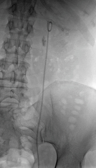
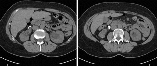
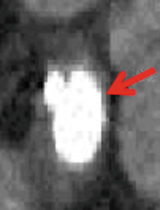
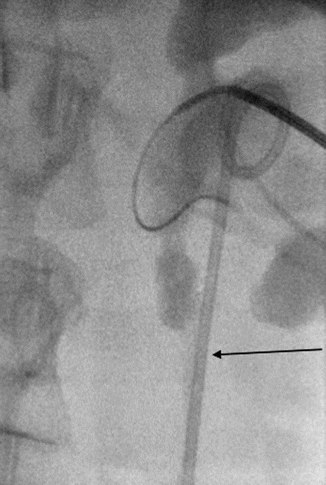
Fig. 13.2
KUB demonstrates significant residual stone burden in the proximal ureter and middle and lower calyces after stenting and three courses of SWL for an impacted ureteral stone. The stent appears to be too far lateral from the stone raising the suspicion for either a perforation or an intramural stone fragment
Fig. 13.3
CT scan confirmed that the proximal coil was outside of the collecting system
Fig. 13.4
Thinning of the ureteral wall associated with stone impaction
Fig. 13.5
A percutaneous antegrade approach was used to clear the impacted stone as well as the renal fragments, with removal of the misplaced ureteral stent
Our Approach to Impacted Ureteral Stones
Preoperative images are reviewed carefully. Duration of symptoms may be one factor that raises the suspicion that a stone may be impacted. On non-contrast CT, on occasion the ureteral wall will appear “thinned,” raising concern for impaction, as will marked hydroureteronephrosis above the stone. If parenchymal thinning is noted, renal scintigraphy may be performed to quantify split renal function.
For stones <10 mm, a retrograde ureteroscopic approach is employed. Initial attempt is made to place a Sensor wire (Boston Scientific, Natick MA) cystoscopically past the point of obstruction. If the wire starts to buckle at the level of the stone, a 5-French open-ended catheter is advanced under fluoroscopic guidance to that point over the Sensor wire and the sensor wire is removed. A gentle retrograde pyelogram is performed to delineate the collecting system. A Glidewire (Terumo) is then advanced through the open-ended catheter and attempts to traverse the point of impaction are made. If the wire will not advance, it may be exchanged for an angled-glidewire. If this fails, 1–2 cc of lubricating jelly is gently instilled through the open-ended catheter and a final attempt at passage of the wire is made.
If the glidewire advances beyond the stone, the open-ended catheter is advanced over the glidewire to the ureteropelvic junction and the wire is removed. Contrast is instilled to confirm no perforation or submucosal false passage. The Sensor wire is then inserted for a more secure safety wire and the 5-Fr open-ended catheter is removed.
If the glidewire does not advance beyond the stone, then a semi-rigid ureteroscope (6Fr Tip) is advanced under direct vision to the level of the stone. A wire may be advanced under direct vision through the scope if a luminal opening is noted beside the stone. If no lumenal opening is identified, the stone can be carefully fragmented with the holmium laser until a lumen is created at which point the safety wire is inserted.
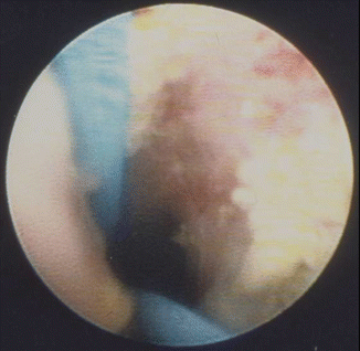
Once the wire is placed, the scope is removed and reinserted alongside the wire, with further fragmentation and clearance of the stone. In the face of an impacted stone, greater care is needed at all steps to avoid perforation. It is critical to clear all small fragments from the point of impaction before advancing the scope or a ureteral access sheath past this point – as failure to do so may lead to fragments embedding in the wall with subsequent stone granuloma and stricture. At times, the fragments may be too adherent to the inflamed mucosa, making extraction impossible (Fig. 13.6). If at any time during the retrograde ureteroscopic approach, the impacted stone cannot be clearly visualized or treated safely and there is no safety wire in place then a nephrostomy tube should be placed. The stone can then be treated via a percutaneous antegrade approach in the future or if an antegrade stent can be placed, a retrograde approach can be re-attempted once the stent has been allowed to passively dilate the ureter.
 Radiation Exposure to the Patient and the Urologist
Shock Wave Lithotripsy for the Treatment of Ureteral Stones
Selecting the Appropriate Treatment Modality for Ureteral Calculi
Complications in the Treatment of Ureteral Stones: Prevention and Management
Radiation Exposure to the Patient and the Urologist
Shock Wave Lithotripsy for the Treatment of Ureteral Stones
Selecting the Appropriate Treatment Modality for Ureteral Calculi
Complications in the Treatment of Ureteral Stones: Prevention and Management
 Semirigid Ureteroscopy for Ureteral Calculi
 Tips and Tricks in the Treatment of Ureteral Stones


Related posts:
Radiation Exposure to the Patient and the Urologist
Shock Wave Lithotripsy for the Treatment of Ureteral Stones
Selecting the Appropriate Treatment Modality for Ureteral Calculi
Complications in the Treatment of Ureteral Stones: Prevention and Management
Semirigid Ureteroscopy for Ureteral Calculi
Tips and Tricks in the Treatment of Ureteral Stones
Stay updated, free articles. Join our Telegram channel
Full access? Get Clinical Tree
