Diarrhoea


Diarrhoea


ACUTE DIARRHOEA

Enriqueta Román Riechmann MD


Definition

Diarrhoea is a change in the individual bowel habit resulting in more frequent and/or looser stools. It expresses an acute gastrointestinal inflammation (acute gastroenteritis). In childhood, gastrointestinal infection is the most common cause of acute diarrhoea worldwide1, 2.


Aetiology

In industrialized countries the most clinically significant agents in infant acute diarrhoea are viruses (Table 3.1), mainly group A rotavirus (3.1A, B). Other viruses involved are human calicivirus (norovirus and sapovirus, formerly known as Norwalk and Sapporo virus), astrovirus and enteric adenovirus (types 40 and 41), with some common features (Table 3.2)3.


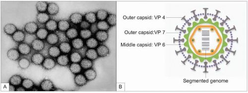
3.1 A: Electron microscopy of rotavirus particles from an infant with acute diarrhoea (courtesy Centers for Disease Prevention and Control, Atlanta, GA, USA); B: schematic of the complete rotavirus particle with structural proteins in the different shells.

Most common bacteria are Campylobacter spp. and Salmonella spp., followed by Shigella, Yersinia and Escherichia coli. The major parasitic infections are Giardia and Cryptosporidium2.


Epidemiology

Viral gastroenteritis is the second most common disease in developed countries. The sporadic form affects all children in the first 5 years of life. Viruses are transmitted fundamentally by the faecal-oral route. There is faecal
excretion of viral particles in the days prior to clinical symptoms and continuing through to its resolution. Rotavirus is a seasonal infection and in temperate climates infections peak during the winter months3. In bacterial enteric infection transmission can be through contaminated water or foodstuffs.


Table 3.1 Main agents of infectious acute diarrhoea


Viruses


Group A rotavirus


Enteric adenovirus


Astrovirus


Human calicivirus


Norovirus


Sapovirus


Parasites


Giardia lamblia


Cryptosporidium parvum


Bacteria


Salmonella


S. typhi and paratyphi


Nontyphoidal Salmonella


S. enteritidis


S. typhimurium


Shigella


Shigella sonnei


Campylobacter


C. jejuni


Yersinia


Y. enterocolítica


Escherichia coli


Enteropathogenic E. coli


Enterotoxigenic E. coli


Enteroinvasive E. coli


Enterohaemorrhagic E. coli


Diffusely adherent E. coli


Enteroaggregative E. coli


Aeromonas


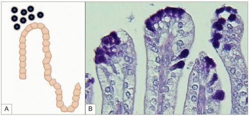
3.2 Pathogenesis of viral diarrhoea: rotavirus infects selectively mature enterocytes on the tips of small intestine villi, leading to their destruction and villi atrophy. (Courtesy Faculty of Biological Sciences, University of Barcelona, Barcelona, Spain.)


Table 3.2 Main features of viral agents


RNA viruses (except adenovirus)


Nonlipoproteic envelope


Seasonal distribution


Asymptomatic infection ↔ severe disease


Frequent coinfections


Endemic, sporadic cases/epidemic, outbreaks


Faecal-oral transmission


Pathophysiology

Diarrhoea occurs when the volume of water and electrolytes present in the colon exceeds its capacity for absorption. This can be mainly due to an increase in the secretion and/or a decrease in the absorption level of the small intestine.

Decreased intestinal absorption occurs as a result of intestinal damage or inflammation (3.2). Viruses causing diarrhoea infect selectively mature enterocytes, causing cell lysis and producing a decrease in disaccharidase activity and in mechanisms for active sodium and water absorption. The consequence is a malabsorptive or osmotic diarrhoea. Diarrhoea caused by bacterial infection is most frequently secretory. Bacteria can activate one of the intracellular pathways leading to intestinal secretion through enterotoxins.


Clinical features

Acute diarrhoea is a self-limiting process. Viral diarrhoea is typically acute in onset, watery-like, and the faeces do not contain mucus, blood, or white cells. Diarrhoea can lead to dehydration, acidosis, and electrolyte imbalance. Vomiting appears at the beginning of the process. The most common age is 6-24 months and rotavirus infection is associated with a more severe disease3.

In the secretory and osmotic diarrhoeas, faeces are watery and profuse. The invasive diarrhoea is frequently characterized by mucus and macroscopic blood. Nevertheless, viral gastroenteritis cannot be distinguished from that caused by bacteria through clinical history or physical examination, although some characteristics may suggest bacterial diarrhoea (Table 3.3).


Table 3.3 Clinical features suggestive of bacterial diarrhoea


  • Children older than 3 years


  • Acute onset


  • No vomiting


  • Hyperthermia


  • Bloody diarrhoea


  • Increase in CRP


  • Faecal white cells


Table 3.4 Assessment of dehydration degree (adapted from ESPGHAN 2001and CDC report 2003)6, 7


Symptom


Minimal or no
dehydration


Mild to moderate
dehydration


Severe dehydration


Body weight loss


<3%


3-9%


>9%


General condition


Well, alert


Restless, irritable


Lethargic or unconscious: floppy


Eyes


Normal


Slightly sunken


Deeply sunken


Tears


Present


Absent


Absent


Mouth and tongue


Moist


Dry


Very dry


Thirst


Drinks normally, not thirsty


Thirsty, eager to drink


Drinks poorly, unable to drink


Skin fold


Instant recoil


Recoil in <2 seconds


Recoil in >2 seconds


Fontanelle


Normal


Sunken


Sunken


Heart rate


Normal


Normal to increased


Tachycardia, bradycardia in most severe cases


Quality of pulses


Normal


Normal or slightly decreased


Moderately decreased


Extremities


Normal capillary refill


Delayed capillary refill


Cool, mottled


Urine output


Slightly decreased


<1 ml/kg/h


<1 ml/kg/h


Assessment

In most cases, a complete clinical history and a careful physical examination is all that is necessary4,5,6. These should rule out any life-threatening cause such as intussusception, surgical abdomen, and haemolityc-uraemic syndrome7. The severity of dehydration is assessed in terms of weight loss as a percentage of total body weight. An assessment of dehydration degree can be made by diverse scales of clinical signs and symptoms (Table 3.4, 3.3).

Supplementary laboratory studies are usually unnecessary. There are some recommendations on which patients should have blood tests (serum electrolytes, urea/creatinine, bicarbonate) (Table 3.5) and on which patients should have faecal laboratory study (Table 3.6), as aetiology is irrelevant for clinical management. Tests for specific pathogens
include stool cultures for bacteria and detection of faecal viral antigen by enzyme immunoassay (EIA), agglutination with latex particles, or immunochromatography.


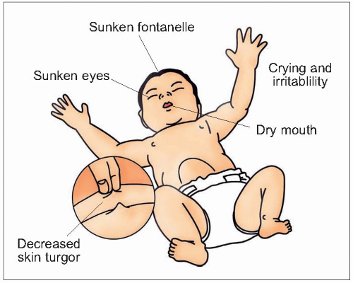
3.3 Signs of dehydration.


Table 3.5 Recommendations on blood tests (adapted from AAP 1996)5, 8


Severe dehydration


Moderate dehydration where clinical signs might indicate hypernatraemia


Moderate dehydrated patients whose histories or physical findings are inconsistent with straightforward diarrhoeal episodes


History of excessive hypertonic or hypotonic fluid ingestion


Table 3.6 Recommendations for faecal laboratory study


Immunocompromised


Blood in the stool


Uncertain diagnosis


Severe or prolonged diarrhoea


Hospitalization


Recent travel abroad


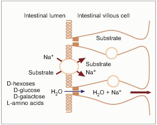
3.4 Diagram to show the processes involved in cotransport of organic solutes and sodium and secondary water absorption.


Treatment

There is no specific treatment for acute gastroenteritis. The main objective is to treat the dehydration and to lead to nutritional recovery. Treatment includes two phases, rehydration with quick replacement of fluid deficit, followed by maintenance in which rapid realimentation and maintenance fluids are indicated.


Rehydration

The molecular process for cotransport of glucose and sodium was the basis for oral rehydration therapy (ORT) development (3.4). ORT is recommended globally for the management of acute diarrhoea2, 6,7,8.

The World Health Organization (WHO), the American Academy of Pediatrics (AAP), and the European Society of Pediatric Gastroenterology, Hepatology, and Nutrition (ESPGHAN) recommend the use of ORT in the treatment of gastroenteritis with mild to moderate dehydration (Table 3.7). Oral rehydration solution (ORS) is more physiological, cost-effective and has fewer adverse effects that intravenous therapy. Intravenous fluids should be reserved for patients with severe dehydration.


Table 3.7 Oral rehydration solution composition


Sodium
(mmol/l)


Potassium
(mmol/l)


Chloride
(mmol/l)


Base
(mmol/l)


Glucose
(mmol/l)


Osmolarity
(mOsm/l)


WHO (1975)


90


20


80


30Bic


110


310


ESPGHAN (1992)


60


20


60


10Cit


74-111


200-250


WHO (2002)


75


20


65


10Cit


75


245


Bic Bicarbonate; Cit Citrate

Only gold members can continue reading. Log In or Register to continue

Stay updated, free articles. Join our Telegram channel

Jun 19, 2016 | Posted by in GASTROENTEROLOGY | Comments Off on Diarrhoea

Full access? Get Clinical Tree

Get Clinical Tree app for offline access