Fig. 10.1
(a, b) CT images showing hydronephrosis of the left kidney due to a distal left ureteric TCC
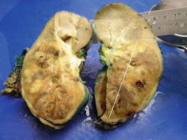
The evaluation of the contralateral kidney is important because of the possibility of bilateral lesions and also for determination of the function in that kidney. Unlike renal cell carcinomas that tend to distort the renal outline, even large infiltrating TCCs tend to maintain a normal renal shape and contour (Fig. 10.2) [2, 3].


Fig. 10.2
Gross pathology of a large TCC infiltrating the left kidney. The tumour involves the lower 3/4 of the kidney, but the outside contour is maintained
Magnetic resonance urography imaging is useful in those who cannot be subjected to CT imaging and has a detection rate of 75 % after contrast injection for tumours <2 cm [4].
Despite limited publication, [18F] Fluorodeoxyglucose PET can be used in UTUC for a better definition of LNs and distant metastases when CT-scan and MRI findings are equivocal.
10.3 Urine Cytology and Cystoscopy
Positive urine cytology is highly suggestive of upper tract urothelial carcinoma when cystoscopy shows normal findings and if carcinoma in situ (CIS) of the bladder is excluded.
10.4 Diagnostic Ureteroscopy
A rigid or flexible ureteroscopy can be performed to obtain a tumour biopsy and determine tumour grade. When the ureteroscopy is unavailable or not feasible, one can also perform selective ureteral cytology and a retrograde pyelogram.
< div class='tao-gold-member'>
Only gold members can continue reading. Log In or Register to continue
Stay updated, free articles. Join our Telegram channel

Full access? Get Clinical Tree


