| 24 | Decompression Tube Placement |
 Definitions
Distention of the colonic lumen has various causes. Two forms of distension are basically distinguished: prevented passage due to mechanical obstruction leading to dilation of proximally located bowel segments and dilation of the colon without mechanical obstruction, i. e., pseudo-obstruction of the colon.
Acute Pseudo-obstruction of the Colon
 Definition and Pathogenesis
In 1948 Leithauser and Ogilvie simultaneously reported acute dilation of the colon without mechanical obstruction (15, 20). This entity, nowadays referred to as acute pseudo-obstruction of the colon (synonym: Ogilvie syndrome) is uncommon, but can lead to massive problems. Radiographs demonstrate distention of the colon, as a rule affecting the cecum, ascending colon, and transverse colon; in rare cases the descending colon is also affected. The decisive factor in diagnosis is distention of the diameter of the cecum to more than 9 cm (Figs. 24.1, 24.2). The cecum in a healthy individual is 3.5–8.5 cm in diameter. Colon haustration is maintained.
Pathogenesis remains somewhat unclear. It is not certain whether primary abnormal motility is in the right or left hemicolon. One possibility is that relaxation of the smooth musculature in the right hemicolon leads to an accumulation of gas and enteral contents. However, it is also possible that abnormal motility in the left hemicolon causes functional obstruction of passage and thus distention of the colon segments proximal to this region. The causes of acute pseudo-obstruction are varied (Tab. 24.1). According to compiled studies (30) pseudo-obstruction occurred in 23% of patients following an operation and in 11% following trauma; in 17% it was associated with cardiopulmonary disease and in 15% with systemic disorders, such as metabolic imbalance, intoxication, and infection.
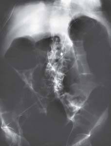
Fig. 24.1 Pseudoobstruction of the colon. Abdominal radiograph in a patient with sepsis syndrome on artificial respiration and with massive distention of the entire colon, especially the cecum (image provided courtesy of Dr. V. Remplik, Institute for Diagnostic Radiology and Neuroradiology, Augsburg Clinic).
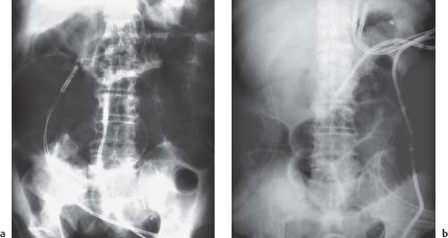
Fig. 24.2 Acute pseudo-obstruction of the colon in an intensive care patient who had to undergo complicated cardiac surgery.
a Abdominal radiograph with distended colon (ECG lead on the abdomen).
b Radiographic image after endoscopic decompression of the colon and placement of a decompression tube (Wilson– Cook), the tip of which is in the transverse colon (images provided courtesy of Dr. V. Remplik, Institute for Diagnostic Radiology and Neuroradiology, Augsburg Clinic).
Postoperative conditions |
|---|
|
Traumatology |
|
Systemic and neurological diseases |
|
Medication/Drugs |
|


 Clinical Picture and Course
The chief symptom is abdominal distention. The abdomen remains soft, however, and does not demonstrate any localized guarding. Around 80% of patients complain of abdominal pain (29), though it is generally not severe. One study reported only 10% of patients complaining of pain symptoms (11). Severe abdominal pain is a sign of complications such as ischemia and perforation. The patient is usually constipated, but may also present with diarrhea or watery stool. Patients complain of nausea and vomiting in slightly more than half of cases.
Perforation and bowel ischemia. In 3% (24) of patients overdistention can lead to perforation of the cecum as the cecum has the thinnest bowel wall of all colon segments. The risk of perforation appears to begin occurring at a cecal diameter > 12 cm; at > 14 cm it rises to 23% (29). Duration of distention of the cecum also appears to be correlated with perforation risk. If perforation occurs, mortality increases to 43–46% (18, 25). In 7–10% of patients with acute pseudo-obstruction of the colon (11, 27, 29), pseudo-obstruction is associated with bowel ischemia, probably as a result of increased transmural pressure. Mortality associated with acute pseudo-obstruction of the colon is difficult to estimate, as most seriously ill patients die of grave underlying causes and not as a result of colon distention. Mortality in conservative therapy is reported at nearly 10% (overview in 30).
 Therapy
Conservative therapy. The first step in therapy of acute pseudoobstruction is the correction of factors supporting it. This includes correcting electrolyte imbalance and other metabolic imbalances. A nasogastric tube should be attached to relieve the upper gastrointestinal tract and the patient should remain on a liquid diet. Forced mobilization and body positioning (frequent changing of patient position, lying on the abdomen with raised pelvis) can help empty the large bowel of gas. The only scientifically confirmedmethod in pharmacological therapy is the use of the cholinesterase inhibitor neostigmine. Ponec et al. (22) showed that in 10 out of 11 patients in the group receiving neostigmine, intravenous administration of 2mg of neostigmine led to a decrease in the diameter of the cecum, stool, and wind; among the 10 patients in the placebo group, there was no effect. However, two of the 10 patients who initially responded to neostigmine suffered a relapse.
Endoscopic therapy. In 1977 Kukora and Dent (14) became the first the show that colonoscopic decompression of the distended bowel in acute pseudo-obstruction is both possible and effective. However, there is a problem in that in 20% of patients successful decompression is followed by recurrence (30). Taking individual reports together, decompression would achieve relief of acute pseudo-obstruction in the colon in 80% of patients (30), although in individual studies such as one by Harig et al. (13
Related posts:


Stay updated, free articles. Join our Telegram channel

Full access? Get Clinical Tree


