This article discusses the evaluation and management of genitourinary system trauma in the critically injured patient. Injuries to any of the organs in the genitourinary system can be managed with a damage control strategy. Often, there is little or no preoperative imaging or injury staging, and these injuries are diagnosed intraoperatively. Finally, specific management strategies of renal, ureteral, bladder, urethral, and genital injuries are discussed.
Key points
- •
Damage control management of urologic injuries is performed in conjunction with the managing trauma team.
- •
Evaluation and management of genitourinary injuries in a damage control setting requires unconventional evaluation and surgical techniques because of the critical nature of this group of injured patients.
- •
Renal injury can often be expectantly managed because of containment of hemorrhage by normal fascial anatomy.
- •
Temporary urinary diversion through externalized tubes and catheters is acceptable and appropriate in the damage control setting.
- •
Complex genitourinary reconstructive surgery should be delayed until the patient is hemodynamically and metabolically stable for prolonged surgery.
Introduction
Damage control in the management of multiorgan trauma is a well-established principal in severely injured patients. The underlying objective is patient survival at all costs. Typically, these patients are critically ill and an abbreviated laparotomy is performed to rapidly control life-threatening bleeding or injuries and the patient is temporarily closed and resuscitated in an intensive care unit setting. The clinical hallmark of patients requiring damage control is the triad of hypothermia, metabolic acidosis, and coagulopathy. Since the term “damage control” was coined in 1993 this multiphase patient management strategy has been expanded to include nonabdominal trauma including thoracic, vascular, and neurosurgical. The genitourinary organ system is also well suited to this style of management in this subgroup of patients.
Over the past 20 years, we have worked to select approaches to management of the urinary system that allow a common patient care philosophy with our trauma general surgeons. Many urologic injuries can be successfully managed with a planned delayed definitive management after initial hemodynamic control and resuscitation including certain renal, ureteral, bladder, urethral, and genital injuries. This article addresses injury selection, initial imaging and injury staging, initial management at damage control, options for definitive management of urologic trauma, and complications of urologic damage control. Additionally, a brief history of damage control and communication and interaction between the urologist the trauma surgeon are discussed.
History of damage control
Damage control is a systematic three-phase approach to management of critically injured patients presenting with the lethal clinical triad. These patients typically undergo an initial abbreviated laparotomy to control life-threatening bleeding and fecal contamination (phase 1). This is followed by fluid resuscitation, correction of coagulopathy, and warming in the intensive care setting (phase 2). Then, after the patient is hemodynamically stable, they are returned to the operating room for definitive management of abdominal injuries and closure of the abdominal wall (phase 3). The evaluation of damage control began in the 1980s with the publication of multiple series evaluating a subset of critical patients with severe abdominal injuries. Before this, the traditional concept was that an injured patient is evaluated in the emergency department and is taken to the operating room for a definitive procedure and the abdomen closed. In the 1980s, Feliciano and Mattox began to evaluate abdominal packing for severe liver injuries. In a series of 300 consecutive abdominal gunshot wounds, they noted that survival in this group was worse in patients who developed the triad of acidosis, hypothermia, and coagulopathy. Four years later, Burch and colleagues described an abbreviated laparotomy in 200 patients with this same triad and noted a survival rate of 33%. The term damage control was coined in 1993 by Rotondo and coworkers after their evaluation of 46 patients, 24 treated with abbreviated laparotomy, and found a survival benefit in patients with significant vascular and multiple visceral injuries. Since that time, damage control has continued to evolve as a major trauma management strategy for critically injured patients and has been applied in the civilian and military settings.
General management issues
Emergency Department
Evaluation of the urinary system for injury has improved over time so that well-defined indications exist for examination, imaging, and laboratory study of these patients. In general, all hemodynamically stable patients with gross hematuria or those patients with blunt or penetrating mechanism, microscopic hematuria, and an episode of hypotension (defined as systolic blood pressure of <90 mm Hg) require contrast-enhanced imaging. In most modern trauma centers the preferred imaging technique is cross-sectional imaging by computed tomography. In those patients who are not hemodynamically stable, imaging studies might not be feasible because often these individuals are rapidly transported to the operating room. Although desirable, confirmation of two functional kidneys preoperatively is not always possible in these settings.
Interaction with the trauma service is beneficial for deciding the urgency and necessity of imaging, facilitating the completion and performance of these studies when necessary, and placing and evaluating urinary drainage catheters. We have found that close preoperative collaboration with the trauma surgery team has facilitated the development of preoperative imaging protocols and streamlined intraoperative evaluation and management of urologic injuries.
Communication
In patients with multisystem trauma, successful management demands frequent and effective communication between various entities, especially between the teams of surgical specialties managing these patients. This is especially true in the damage control patient because their overall survival is lower as a result of the frequent triad of hypotension, hypothermia, and coagulopathy.
The optimal time for consultation of surgical subspecialties by the trauma surgery service is “as soon as possible,” irrespective of need for initial support. In our hospital, we are routinely notified in instances of penetrating trauma when the patient has gross hematuria while the patient is still being evaluated in the shock room. When notified at this stage, we can participate in the entire sequence of urinary system evaluation and management from initial recommendations for radiologic studies through possible surgical treatment. Furthermore, we are able to rapidly assist with drainage or diversion of the urinary system and discuss expected outcomes caused by delaying definitive management of the urinary system injuries. Although the trauma service ultimately controls patient management decisions, we believe that our early input allows for more complete patient care and improvement in outcomes in these critically injured patients.
Intraoperative Evaluation
In patients taken emergently to the operating room multiple options exist for evaluation. The “one-shot” intravenous urogram performed in the operating room provides basic information before intervention, namely the presence or absence of bilateral nephrograms. One caveat in this setting is that a hypotensive patient may not have the intravascular volume or pressure to filter enough contrast through the collecting system to create a nephrogram. A second, less conventional, option is to administer a vital dye, such as methylene blue or indigo carmine, intravenously and atraumatically occlude the ureter of the injured kidney. Contralateral function can be confirmed with the collection of discolored urine in the catheter collection bag.
Injuries to the lower urinary tract should not delay urgent intervention when ongoing blood loss creates concern for patient survival. Evaluation of the urethra and bladder can readily be performed in the operating room after significant hemorrhage has been controlled. In patients with suspected urethral injury, options for intraoperative evaluation include cystoscopy or retrograde urethrogram after abdominal exploration. These evaluations should be performed before catheter insertion. The bladder can be evaluated by exploration and bladder distention with retrograde infusion of vital dye or with static stress cystogram after abdominal exploration and control of hemorrhage.
External genital injury is rarely life threatening and does not require preoperative evaluation in the unstable patient. Key considerations are control of hemorrhage with pressure dressing or clamping and suture ligation of bleeding vessels intraoperatively. Hemorrhage control can be performed either concomitantly with abdominal exploration or after life-threatening issues have been addressed.
Intraoperative Hemorrhage
Significant sources of bleeding within the genitourinary system include kidney, bladder, urethra, and genitalia. The specific management of organ-specific injury is discussed later; however, several important considerations merit special mention. Typically, retroperitoneal bleeding in the kidney is confined within Gerota’s fascia or the perirenal fascia and can be left undisturbed. Indications for exploration include pulsatile retroperitoneal hematoma or ongoing bleeding through or around the perirenal fascia. Bladder hemorrhage manifests in two forms: extravesical or intravesical. Extravesical hemorrhage can occur into either the pelvis or abdominal cavity by laceration of the perivesical blood vessels or the detrusor muscle. Intravesical hemorrhage occurs from mucosal or detrusor laceration and can cause rapid, profuse gross hematuria. With extravesical hemorrhage, rapid suture ligation of bleeding or perivesical packing with laparotomy pads for control may be necessary. For intravesical hemorrhage, temporary hemorrhage control can be facilitated by catheter occlusion and clot tamponade with correction or repair after the patient is stable. Genital bleeding is usually controlled with suture ligation, but definitive complex genital reconstruction often requires a future trip to the operating room.
Uncontrolled Urinary Extravasation
Although desirable, early urinary control is important but lower priority in the order of patient management issues. In contrast to uncontrolled fecal contamination, which can result in early sepsis, urine is typically sterile and does not pose this risk. However, ongoing urinary extravasation does create tissue irritation and can cause perinephric, retroperitoneal, peritoneal, or pelvic inflammation. Because many techniques of temporary abdominal closure involve vacuum dressing or closed suction drains, often much of the extravasated urine is removed. Additionally, many options for temporary urinary diversion exist that can be rapidly implemented on either the initial exploration or at future surgery. This allows for definitive repair to be delayed or deferred until the patient is hemodynamically stable and reconstructive surgery is appropriate. For upper-tract injuries, an externalized stent can be quickly placed and provide thorough urinary diversion. In lower-tract injuries, such as significant bladder injuries, which cannot be quickly repaired, ureteral diversion stents prevent or lessen pelvic or abdominal urinary extravasation and subsequent inflammatory response.
Management of specific organ injuries
Renal Injury
Our approach to intraoperative evaluation and management of renal injuries begins with an assessment of the patient’s hemodynamic stability and discussion with the operating trauma service about management goals. Often, a patient undergoing abbreviated laparotomy and damage control management has limited preoperative radiologic evaluation. In these cases, decision-making is based on operative findings and available adjunctive studies. Most renal injuries are amenable to a damage control approach with the exceptions being expanding or pulsatile hematoma and ongoing uncontained hemorrhage. We have found that expectant management is successful and appropriate in those patients who are not hemodynamically stable enough to undergo the necessary reconstruction in salvageable injuries. Additionally, it is common practice in urologic trauma patient management to observe and monitor high-grade renal injuries from blunt mechanism. Our approach in damage control patients is to evaluate but not necessarily explore a confined retroperitoneal hematoma or hemorrhage. Delayed formal exploration and reconstruction can then be performed in a fully resuscitated and more stable patient.
In those patients with penetrating injuries, the injury is often apparent based on the site of retroperitoneal penetration. Blunt injuries can be more challenging because the retroperitoneal fascia is often intact and without the benefit of preoperative imaging, the exact location of the injury is more difficult to ascertain. In these patients, evaluation of the hematoma is paramount in deciding which patients to explore. We have observed that retroperitoneal hematomas requiring exploration are relatively easy to identify, usually indicate renal pedicle injury, and are a life-threatening emergency. The most difficult patients to manage are those with an intimal arterial disruption causing vascular occlusion. In those patients without preoperative imaging, the surgeon must decide if the injury warrants exploration and if the patient is stable enough to undergo evaluation and repair of the arterial injury. Often, in patients undergoing abbreviated laparotomy, the patient’s condition dictates that unilateral arterial intimal repair be delayed, especially in the presence of a normal contralateral kidney. In these cases, prolonged warm ischemia usually results in irreparable damage and renal loss.
Further retroperitoneal exploration can be performed without disturbing the contained renal hematoma. Central retroperitoneal hematomas (zone 1) are concerning for vascular injuries, namely the aorta and vena cava. If an injury to these structures is suspected, the trauma surgeon can enter the retroperitoneum and leave the confined renal hematoma undisturbed within the perinephric fascia. If the perirenal fascia is violated and renal cortical bleeding is noted, packing the renal fossa tightly with laparotomy pads can salvage the kidney in an unstable patient. At a future operation, the packs can be removed, the wound irrigated, and the injury repaired in the appropriately selected patient.
A risk of damage control management is that ongoing or even life-threating hemorrhage can occur from the injured kidney while the patient is resuscitated, warmed, and awaits re-exploration and repair. However, our experience has shown that this is not a frequent occurrence and careful monitoring, as in all patients undergoing damage control, is sufficient. In the damage control setting, major renal parenchymal injury can result in urinary extravasation that is difficult to control without significant reconstructive efforts. Our usual approach is to begin with external drainage and plan for management at a future operation. In general, a brief period of controlled local urinary extravasation is unlikely to result in a significant adverse event or impact overall recovery of the patient.
During phase 2 in the damage control process, which is usually a period of 48 to 72 hours, suspected renal injuries identified in the first laparotomy are staged. In most patients, we obtain cross-sectional imaging with contrast-enhanced computed tomography scans to identify injuries and select patients for subsequent operation and reconstruction or continued expectant management with no plan for operative intervention. Identifying renal injuries that are appropriate for nonoperative management allows the trauma surgery team to concentrate on other critical injuries and does not expose the patient to the potential morbidity of unnecessary renal surgery. The most important point is to fully stage unknown or suspected injuries.
In select patients, adjunct imaging including arteriography may be appropriate. In these situations, arteriography can be diagnostic and therapeutic for renal, vascular, or solid organ injury. In some injured patients, angioembolization is an appropriate and less morbid treatment especially during phase 2 of damage control management. However, emerging data show an overuse of angioembolization in the management of renal injury based on grade of the injury. An individualized approach to the selection of operative versus angiographic control of delayed renal hemorrhage is important and should be based on factors including level of radiologic expertise available, other nonurologic indications, severity of renal injury, and stability of the patient.
In patients who require delayed definitive operative management, the concepts and operative steps are similar to other renal trauma management. We believe that initial hilar control is important and thus the hilum is initially accessed to provide for arterial and venous control. We have also developed an alternative method of vascular control that does not involve individual control of the renal artery and vein. In this technique, vascular control is obtained by bluntly dissecting along the plane of the psoas muscle fascia, adjacent to the great vessels, and directly placing a vascular pedicle clamp on the renal hilum. We have found this particularly helpful in instances where significant or rapid bleeding is noted. Regardless of the method of vascular control used, it is important to avoid unnecessary nephrectomy because of diffuse bleeding from inadequate control of the renal hilum. After vascular control, the kidney is mobilized, the perirenal fascia opened, and the hematoma is evacuated. After the kidney is adequately exposed, the injury is debrided and definitive repair is performed. We believe that ureteral stenting or nephrostomy diversion should be strongly considered after delayed reconstruction because of the increased risk of postoperative urinary extravasation compared with primary repairs.
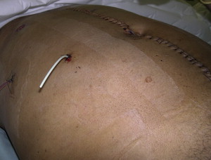
Ureteral Injury
Damage control management of ureteral injury is readily applicable for many reasons. Primarily, the risk of bleeding secondary to a ureteral injury is minimal and outweighed by more pressing concerns with other injuries. Additionally, there are many available options for urinary diversion which can be rapidly implemented that preserve tissue quality and anatomic relationships. Finally, ureteral reconstruction can be a challenging and time-consuming endeavor. Depending on the level of ureteral injury and the status of surrounding tissues, temporary urinary diversion or adjuvant measures may be the best management option when the patient is unstable and resuscitation in the intensive care unit is necessary.
A ureteral injury can be diverted using any number of small, hollow medical tubes to intubate the ureteral lumen and externalize the open end of the selected tube. Our preference is to use “single-J” stents ( Fig. 1 ), commonly used in elective urinary diversion procedures; however, a Cordis catheter, central line, or feeding tube can be used with equal efficacy if a specific stent is not available. Regardless of diversion method, it is important to use a guide wire to ensure that the device travels proximally into the renal pelvis. We secure the stent or tube to the proximal ureter using a 3-0 silk suture and only compress a minimal amount of ureteral tissue (1–2 mm). This prevents migration or dislodgement of the stent and at the same time preserves ureteral tissue for future repair. The distal end of the stent is externalized through a stab incision in the abdominal wall and secured to the skin, after which a drainage bag is placed. With the urine diverted in this fashion, renal output can be monitored until the patient can safely return to the operating room for definitive repair. If the stent becomes occluded with blood clots or debris, the free lumen can be gently irrigated with saline until drainage resumes. If there is any question as to the position of the stent, either plain radiographs or injection of contrast confirms the location of the proximal aspect of the stent. In cases of complete ureteral transection, we do not manipulate, ligate, or divert the distal end of the ureter. Usually, urinary reflux is not a concern in injured patients. If the distal ureter is manipulated or ligated this damages the tissue resulting in ureteral tissue loss and affects the future reconstruction of the ureter.