CT Urography Pitfalls and Artifacts
Richard H. Cohan MD
Elaine M. Caoili MD
History: 55-year-old woman with intermittent hematuria.
Case 11-1
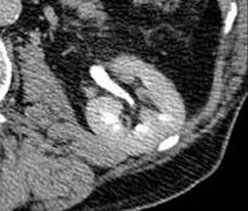
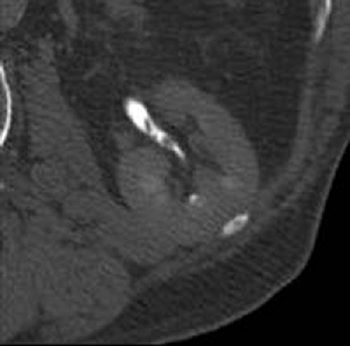
Findings: Axial excretory phase image (A), viewed utilizing standard soft tissue windowing, demonstrates a nondistended left renal collecting system. No masses are identified and there is no urothelial wall thickening. In comparison, when the same image is assessed with wider windows (B), three distinct filling defects, each measuring 5 mm or less, can be identified.
Diagnosis: Multifocal transitional cell carcinoma
Potential Pitfall: Urinary tract pathology will be missed if images are reviewed using only standard soft tissue windowing.
Discussion: Due to volume averaging with high-attenuation excreted contrast material, some intrarenal collecting system, ureteral, and bladder abnormalities will not be detectable if images are reviewed using only standard soft tissue windowing. This problem is more likely when the pathology is small or subtle, as in this case. In order to avoid this pitfall, it is essential that excretory phase images be reviewed utilizing wide windows (often set between 2,000 and 4,000 HU). In this patient, if the left intrarenal collecting system were viewed only with standard soft tissue windowing, it would have been interpreted erroneously as normal. Each of the tiny filling defects was subsequently determined to be a focus of transitional cell carcinoma (see Cases 10-4 and 10-5).
Case 11-2
History: 47-year-old male with recurrent obstructing ureteral calculi and urinary tract infection.
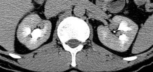
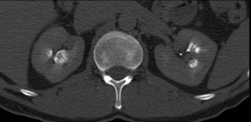
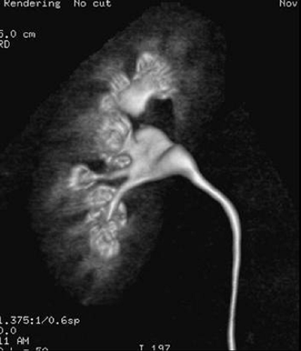
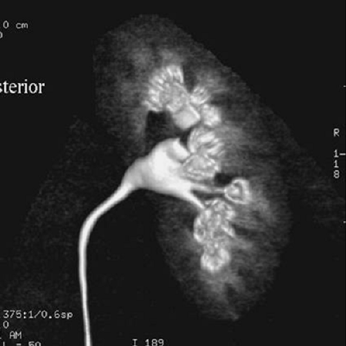
Findings: Excretory phase axial image from a CT urogram (A), viewed at soft tissue windows, demonstrates prominent calices bilaterally. Some of the visualized centrally located high-attenuation contrast material may actually reside in the renal pyramids; however, when using soft tissue windows the appearance is most consistent with a prominent papillary blush. In comparison, when the same image is viewed with wide windows (B), discrete linear rays of contrast-enhanced urine are identified in the renal pyramids. Similar findings are demonstrated in both kidneys on the volume-rendered images (C, D).
Diagnosis: Renal tubular ectasia
Potential Pitfall: Urinary tract pathology will be missed if images are reviewed using only standard soft tissue windowing.
Discussion: Occasional intrarenal collecting system, ureteral, and bladder abnormalities will not be detectable if images are reviewed using only standard soft tissue windowing. This problem is more likely when the pathology is small or subtle, as in this case. In order to avoid this pitfall, it is essential that excretory phase images be reviewed utilizing wide windows. In this patient, the discrete linear collections of opacified urine are identified only on images viewed with wide windows. The findings are diagnostic of renal tubular ectasia (see Cases 4-10 and 8-1).
Case 11-3
History: 73-year-old male with intermittent microscopic hematuria.
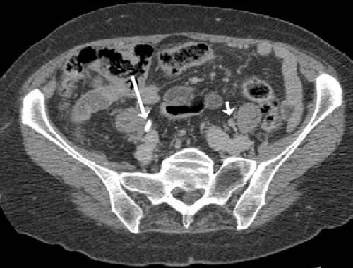
Findings: Two excretory phase axial images (A, B) demonstrate opacification of the right (long arrow) but not the left ureter (short arrow); however, both ureters can be seen to be normal in caliber.
Diagnosis: Left ureteral polyp
Potential Pitfall: Nonopacification of a nondilated ureter preventing detection of pathology.
Discussion: There are three reasons why urinary tract segments may not be opacified on CT urograms: (i) obstruction resulting in delayed excretion into usually dilated renal collecting systems and ureters; (ii) peristalsis in normal nondilated ureters; and (iii) layering of excreted contrast material in dilated portions of the renal collecting systems, ureters, and urinary bladder. Intrarenal collecting system and ureteral pathology is usually detectable when present in the dilated urinary tract. In particular, the cause of obstruction is frequently identified. In comparison, tiny abnormalities in unopacified nondistended portions of the intrarenal collecting systems and ureters may be impossible to detect, as in this case, in which a small left ureteral polyp was later diagnosed at the level illustrated here.
There are a number of possible approaches to the problem of unopacified nondilated urinary tract segments. Some radiologists obtain additional, more delayed, axial CT images, whereas others obtain delayed digital scout (or scan projection) radiographs. Still others perform no additional imaging and report these segments as normal, with the understanding that in rare instances, such as in the case illustrated here, abnormalities will be missed.
Case 11-4
History: Elderly male with gross hematuria and negative cystoscopy.
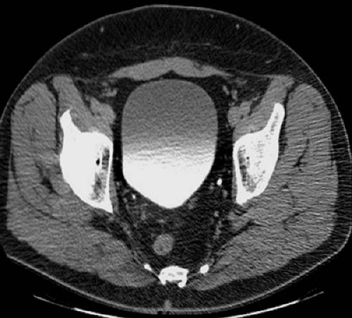
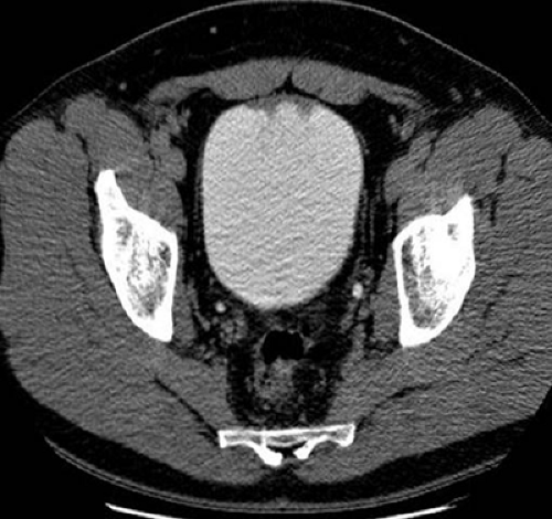
Findings: Excretory phase axial CT urography image obtained with the patient supine (A) demonstrates layering of opacified urine in the bladder. The bladder wall is not thickened and no intraluminal filling defects are identified. The second image (B), obtained several minutes later, after the patient was removed from the scanner and ambulated, shows that the contrast material and urine are now evenly mixed in the bladder. Irregular thickening along the anterior bladder wall is now apparent.
Diagnosis: Transitional cell carcinoma of the bladder
Potential Pitfall: Incomplete opacification of the bladder may prevent detection of bladder wall or luminal abnormalities.
Discussion: If the patient is kept in the supine position after contrast material excretion begins, urine containing contrast material will layer posteriorly in the bladder. Although many bladder lesions in the anterior aspect of the bladder can still be detected in this circumstance (outlined by lower water attenuation unopacified urine), this layering phenomenon may also result in failure to detect an occasional bladder lesion. This pitfall is particularly problematic in patients such as this, who can have normal cystoscopy. Indeed, the two blind spots during flexible cystoscopy are along the anterior wall of the bladder and at the bladder base. It is particularly important that these two areas be optimally imaged during CT urography. In order to do this, several measures may be taken to minimize the amount of unopacified urine in the bladder. Patients can be asked to void completely prior to CT urography. Alternatively, patients can be asked to roll over on the CT table several times and/or to ambulate just prior to excretory phase image acquisition. Others are willing to accept a slight decrease in study sensitivity. Without these maneuvers, CT urography technique is more straightforward and more simply performed because the patient can remain stationary. (Case courtesy of Nigel C. Cowan, Oxford University, Oxford, UK.)
Case 11-5
History: 75-year-old female with hematuria.
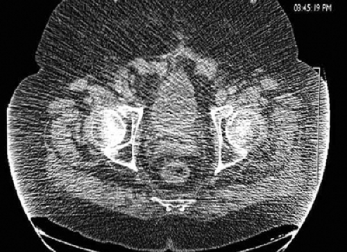
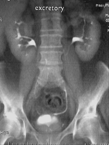
Findings: Single excretory phase axial image (A) has been obtained prior to excretion of injected contrast material into the bladder. The bladder contour is lobulated; however, the bladder wall is not markedly thickened. Average intensity projection image (B) demonstrates apparent wall thickening at the bladder base. Both images have a “grainy” appearance. A coronal excretory phase average intensity projection image obtained several minutes later demonstrates a small amount of contrast material in the bladder.
Diagnosis: Transitional cell carcinoma
Potential Pitfall: Reduced signal-to-noise ratio in large patients limits evaluation of the bladder.
Discussion: This patient is large. The increased soft tissue produces more x-ray beam scatter and there is a reduced signal-to-noise ratio. Generated axial images appear grainy and are of limited quality. This likely contributed to the inability to detect an 8-cm bladder neoplasm, that was subsequently identified at cystoscopy, in this patient.
Case 11-6
History: Elderly patient with gross hematuria.
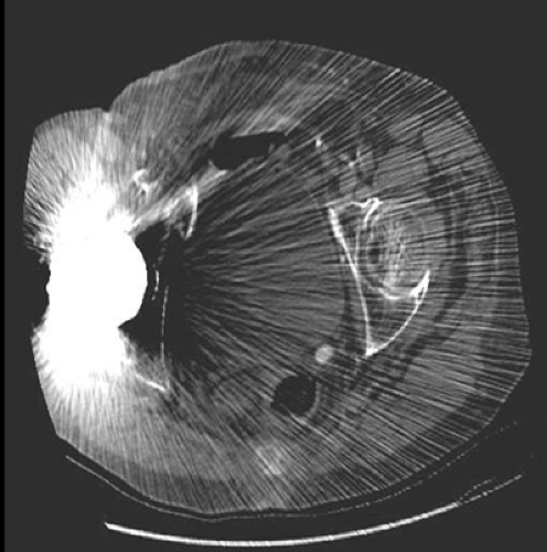
Findings: Excretory phase axial image demonstrates extensive artifact from a metallic right hip prosthesis. This limits visualization of nearly the entire pelvis, but particularly the right side of the bladder.
Diagnosis: Transitional cell carcinoma
Stay updated, free articles. Join our Telegram channel

Full access? Get Clinical Tree


