Study or subgroup
Positive CE
Positive DBE
DBE yield after positive CE
DBE yield after negative CE
Matsumoto et al. [63]
10/13 (76.9 %)
6/13 (53.8 %)
–
–
Hadithi et al. [34]
28/35 (80 %)
21/35 (60 %)
20/28 (71.5 %)
1/7 (14.3 %)
Mehdizadeh et al. [12]
63/115 (54.8 %)
57/115 (49.6 %)
41/63 (65.1 %)
16/52 (30.8 %)
Nakamura et al. [33]
17/28(60.7 %)
12/28 (42.9 %)
9/17 (52.9 %)
3/11 (27.3 %)
Fujimori et al. [32]
18/45 (40 %)
18/36 (50 %)
16/16 (100 %)
2/20 (10 %)
Ohmiya et al. [31]
37/74 (50 %)
39/74 (52.7 %)
–
–
Kameda et al. [30]
23 (71.9 %)
21 (65.6 %)
15/23 (65.2 %)
2/3 (66.7 %)
Arakawa et al. [42]
40/74 (54.1 %)
40/74 (63.5 %)
36/40 (90 %)
11/34 (32.4 %)
Fukumoto et al. [29]
16/42 (38.1 %)
18/42 (42.9 %)
–
–
Marmo et al. [44]
174/193 (90.2 %)
132/193 (68.4 %)
124/174 (71.3 %)
8/19 (42.1 %)
Shisido et al. [27]
53/118 (44.9 %)
63/118 (53.4 %)
51/53 (96.2 %)
12/65 (18.5 %)
A meta-analysis of 10 studies comparing capsule endoscopy with double-balloon enteroscopy yielded a diagnosis rate of 62 % for capsule endoscopy (95 % confidence interval [CI], 47.3–76.1 %) and of 56 % for double-balloon enteroscopy (95 % CI, 48.9–62.1 %) with an odds ratio (OR) of 1.39 (95 % CI, 0.88–2.20, p = 0.61) [28]. The diagnosis rate of double-balloon enteroscopy following capsule endoscopy was 75.0 %, whereas the diagnosis rate of double-balloon enteroscopy without capsule endoscopy was 27.5 %.
According to the study by the Korean Gut Image Study Group, the diagnosis rates for capsule endoscopy and double-balloon enteroscopy in cases of OGIB are similar [35]. However, no randomized controlled studies of the two procedures for OGIB have been undertaken. A meta-analysis of six prospective studies and three retrospective studies showed that the pooled OR for the diagnostic yield was 1.48 (95 % CI, 0.90–2.43). No difference was noted between the two procedures. In patients with OGIB , OR between double-balloon enteroscopy after positive result of capsule endoscopy and double-balloon enteroscopy from the beginning was 1.79 (95 % CI, 1.09–2.96) in a meta-analysis [35]. Generally, capsule endoscopy is more economical than double-balloon enteroscopy. The use of double-balloon enteroscopy based on the capsule endoscopy result is then cost-effective [36].
Particularly, the ability of double-balloon enteroscopy to check the entire small intestine is important in patients with OGIB. Capsule endoscopy facilitates observation of the small intestine without discomfort in 79–90 %. In contrast, the rate to observe the entire small intestine in the double-balloon enteroscopy is reported as 16–86 % [37, 38]. Technical difficulties prevent the observation of the entire intestine using double-balloon enteroscopy . If the cause of bleeding is detected during the procedure, the procedure might not be performed to completion. According to three meta-analyses, the diagnosis rate of double-balloon enteroscopy for OGIB was approximately 60 %, similar to that of capsule endoscopy [13, 28, 39]. Compared to single-insertion (per-anal or per-oral) double-balloon enteroscopy , capsule endoscopy was superior, whereas compared to double-insertion (per-anal and per-oral) double-balloon enteroscopy , capsule endoscopy was comparable [39]. In addition, small bowel lesions were more likely to be found when double-balloon enteroscopy was performed soon after the bleeding episode (for example, <1 month later) as was the case for capsule endoscopy [40, 41].
According to Shishido and colleagues, a total of 118 patients underwent capsule endoscopy and then underwent double-balloon enteroscopy via both anal and oral approaches [27]. The overall diagnosis rates of capsule endoscopy and double-balloon enteroscopy were 44.9 % in 53 patients and 53.4 % in 63 patients (p = 0.01), respectively, with good agreement between the two procedures (kappa statistic = 0.76). In a study comparing double-balloon enteroscopy and capsule endoscopy in 162 patients with OGIB, double-balloon enteroscopy was superior to capsule endoscopy for the detection of lesions in the Roux-en-Y loop or diverticular disease [42].
Based on many studies, the relationship between capsule endoscopy and double-balloon enteroscopy at least in cases of OGIB seems to be complementary rather than competitive [43, 44]. Double-balloon enteroscopy has the advantage of therapeutic procedure in the cases of persistent bleeding or when the bleeding site is confirmed during capsule endoscopy in OGIB. Bleeding in the small intestine can be treated using various hemostasis methods such as injection therapy, argon plasma coagulation, and hemoclipping.
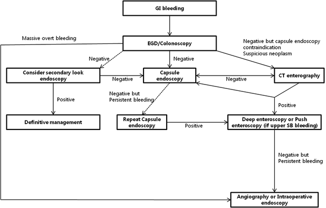
Many clinical guidelines suggest that a combination of the two endoscopy techniques be used [45–47]. The guidelines also suggest that double-balloon enteroscopy be performed after capsule endoscopy. This makes it possible to select the most efficient approach (per-oral or per-anal) [48, 49]. The use of double-balloon enteroscopy immediately after standard endoscopy is recommended for the diagnosis and treatment of acute massive bleeding. Based on the above results, a flowchart for the diagnosis and management of OGIB is shown in Fig. 8.1.
8.1.3.2 Small Intestine Crohn’s Disease
Capsule endoscopy has a very high diagnosis rate in patients already diagnosed with or suspected to have Crohn’s disease. For Crohn’s disease confined to the small intestine, the diagnosis rate of capsule endoscopy was superior (incremental yield = 38 %) to that of push enteroscopy [50]. It was also superior to abdominal computed tomography (CT). Another study also showed that capsule endoscopy has a higher diagnosis rate than other procedures for Crohn’s disease confined to the small intestine [51]. A meta-analysis comparing the diagnosis rates between capsule endoscopy and double-balloon enteroscopy showed no difference between the two. However, no study to date has compared the two procedures for suspected Crohn’s disease . According to a meta-analysis of 11 studies consisting of 375 patients with suspected small intestine lesions that compared capsule endoscopy and double-balloon enteroscopy , the detection rates of an inflammatory disease of the small intestine were comparable (pooled yield 16 % with double-balloon enteroscopy and 18 % with capsule endoscopy) [13].
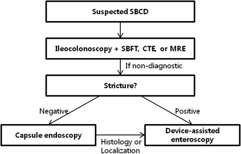
Capsule endoscopy is less invasive than balloon enteroscopy and is more likely to facilitate observation of the entire small intestine. Additionally, capsule endoscopy is generally less expensive and less difficult than balloon enteroscopy. In fact, the results of capsule endoscopy can help determine the approach that should be used for balloon enteroscopy. Thus, the use of capsule endoscopy rather than balloon endoscopy is preferred in cases of suspected Crohn’s disease [45]. However, capsule endoscopy has two limitations. First, a biopsy cannot be performed. Second, the capsule may be retained within the intestine. The incidence of a capsule remaining in the intestine of healthy subjects and patients with inflammatory bowel disease was 1–6 and 7–13 %, respectively (Table 8.2) [13, 38]. Thus, the presence of stricture in patients with suspected or established Crohn’s disease requires confirmation using small bowel series, CT enterography, or MR enterography and then capsule endoscopy should be performed. When capsule enteroscopy or radiological imaging studies reveal positive or suspected results, device-assisted enteroscopy may be conducted to perform a biopsy. Additionally, in cases of clinically suspected small intestine stricture, device-assisted enteroscopy is more useful for diagnostic and treatment purposes, and the stagnant capsule endoscope due to stricture associated with Crohn’s disease can be removed.
Capsule endoscopy | Double-balloon enteroscopy | |
|---|---|---|
Sedation requirement | None | Yes |
Evaluation of the entire SB | 79–90 % | 16–86 % |
Diagnostic yield of small bowel disease | 60 % | 57 % |
Risks | Capsule retention , overall 1–6 %, CD 7–13 % | Pancreatitis 1 % Perforation 1 % |
In particular, the availability of balloon enteroscopy in patients already diagnosed with Crohn’s disease places greater emphasis on treatment. In a prospective study, 11 patients with small bowel strictures due to Crohn’s disease underwent balloon angioplasty via balloon enteroscopy. As a result, the patients’ subjective symptoms improved significantly despite the fact that only a small number of patients were studied [52]. However, balloon enteroscopy is unlikely to facilitate examination of the entire small intestine compared to imaging studies or capsule endoscopy . The disadvantages of balloon enteroscopy also include patient discomfort due to the long procedure time, high cost, and the risk of complication s such as perforation or pancreatitis.
Capsule endoscopy and device-assisted enteroscopy are used in cases of Crohn’s disease as shown in the flowchart in Fig. 8.2.


Fig. 8.2
Flowchart for the diagnosis of Crohn’s disease . SBCD small bowel Crohn’s disease, SBFT small bowel follow through, CTE computed tomography enterography, MRE magnetic resonance enterography
8.1.3.3 Small Intestine Tumors
The incidence of primary tumors in the small intestine is very low, accounting for approximately 3–6 % of gastrointestinal tumors and 1–3 % of malignant gastrointestinal tumors [53–56]. The proportion of malignant small intestine cancers among all malignant cancers is 0.3 %. Overall, cancer of the small intestine is rare. Reasons for this are related to the transit time of the contents of the small intestine and the high levels of enzymes that break down carcinogens [56]. Well-developed lymphoid tissues secrete large amounts of immunoglobulin A, while the alkaline digestive fluid inhibits the propagation of bacteria. Additionally, the removal and regeneration cycles of epithelial cells in the intestinal mucosa are very short [57]. The perceived rarity of tumors of the small intestine could be attributable to the lack of appropriate diagnostic tests. However, in studies using capsule endoscopy and enteroscopy that enabled direct observation of the lumen of the small intestine, the incidence of small intestine tumor s was 2.4–4.3 % in South Korea and Europe [58, 59]. This finding suggests the possibility that the actual prevalence of small intestine tumors is higher than suspected.
The diagnosis rate of capsule endoscopy for small intestine tumors in the entire population has not yet been conducted. In a retrospective study based on a registry of cases in which capsule endoscopy of the small intestine was used for a variety of clinical indication s, the reported diagnosis rate was approximately 4 % [58, 59].
A recent multicenter study in South Korea analyzed the usefulness of the capsule endoscopy in cases limited to small intestine tumor s. In that study, 12.3 % of the small intestine tumors identified by the capsule were malignant and were then surgically resected [58]. The overall diagnostic efficiency of balloon enteroscopy in patients with OGIB was 50.0–61.1 %, treatment was possible in 27.8–35.0 %, and the frequency of diagnosed small intestine polyps or tumors was 6–10 % [60]. In a single study on OGIB, the overall diagnostic efficiency reached 45 % and tumors of the small intestine were found in 13 % of cases [61].
In South Korea, cases of all double-balloon enteroscopy procedures were analyzed regardless of indication . In that study, small intestine tumor s were found in 13.8 % of cases, similar to the 7–20 % rate reported in studies of other countries [62]. In a study compared double-balloon enteroscopy and capsule endoscopy in nine patients with GI polyposis, double-balloon enteroscopy detected a larger number of polyps than capsule endoscopy [63]. The frequency of the tumor diagnosis with double-balloon enteroscopy was relatively high compared to that with capsule endoscopy (4 %). The main reason for this is that the balloon endoscope was actively manipulated to obtain images of the intestinal lumen and is usually preformed after the other tests compared to the capsule endoscope. The possibility of operator manipulation lends an advantage to enteroscopy over capsule endoscopy for diagnosing tumors of the small intestine.
A total of 18 cases diagnosed using double-balloon enteroscopy were analyzed, one-third of which were detected by capsule endoscopy , whereas four cases of small intestine adenocarcinoma s were missed [64]. Double-balloon enteroscopy is useful for the diagnosis and treatment of patients with familial adenomatous polyposis FAP and Peutz-Jegher syndrome (PJS ), groups at high risk of developing adenomatous polyps in the small intestine [65, 66].
Deep small bowel enteroscopy is useful in the diagnosis of causative diseases by facilitating observation and biopsy of neoplastic lesions in the small intestine. Further, this procedure can help perform surgical treatment through impacting the extent of surgery by enabling submucosal injection in the area of the lesion and disclosing the lesion’s exact location [67, 68]. In addition, if hemorrhage associated with the tumor is detected, hemostasis is possible. Deep enteroscopy is also a useful therapeutic tool in the treatment of polyps [69].
8.2 Conclusion
A variety of enteroscopy methods have been developed to diagnose and treat diseases of the small intestine. Of these, the application of capsule endoscopy and balloon enteroscopy has greatly impacted the diagnosis and treatment of small intestine diseases that were previously difficult. According to the results of several studies, these two procedures are able to improve the diagnosis rate of diseases of the small intestine, affect the clinical course and treatment, and play a complementary role in the their management.
Capsule endoscopy is likely to be non-invasive and enables the observation of the entire small intestine. As such, it can be used in cases in which common endoscopic examination is difficult. Balloon enteroscopy is available for various purposes in addition to diagnosis, such as tissue biopsy and endoscopic therapy. Double- and single-balloon enteroscopy are currently being used in clinical practice and do not differ significantly in terms of lesion observation rates and success rates. Of the 2 techniques, double-balloon enteroscopy is considered deep small bowel endoscopy that can be selected to examine the entire small intestine given that it has the highest observation rate of the entire small intestine.
Once we understand the complementary roles of these two endoscopies and apply them in clinical practice, management of small intestinal diseases can be expected to improve.
References
1.
Kwon JG. Indications and choice of small bowel endoscopy. The 48th Korean Society of gastrointestinal endoscopy seminar. 2013. p. 216–220.
2.
Bang S, Park JY, Jeong S, Kim YH, Shim HB, Kim TS, Lee DH, Song SY. First clinical trial of the “MiRo” capsule endoscope by using a novel transmission technology: electric-field propagation. Gastrointest Endosc. 2009;69:253–9. doi:10.1016/j.gie.2008.04.033 (PMID: 18640676).PubMedCrossRef
Stay updated, free articles. Join our Telegram channel

Full access? Get Clinical Tree


