Fig. 14.1
The epithelium alone is dissected away from the underlying fibromusclar layer of the vaginal wall (a, b). (Photo credit: Dr. Marlene Corton, with permission)

Fig. 14.2
The vaginal tube with excised epithelium has been everted. Successive purse-string sutures will begin closest to the vaginal apex, which is pushed inward with subsequent rings of suture.(Photo credit: Dr. Marlene Corton, with permission)
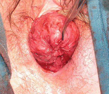
Fig. 14.3
The vaginal tube continues to be elevated with resolution of the prolapse by pushing inward/cephalad with a hemostat or similar instrument on each successive suture ring. (Photo credit: Dr. Marlene Corton, with permission)
Partial (LeFort) Colpocleisis
As above, the vaginal apex (or cervix) is grasped with clamps and delivered as far outside the introitus as possible (Fig. 14.4). Rectangular areas of approximately equal dimensions are delineated with either marker or cautery on both the anterior and posterior vaginal walls. The hashed blue lines in Fig. 14.4 delineate the intended region for dissection and skin excision on the anterior vagina. The length of the vaginal walls ultimately dictates the rectangles’ size, but generally the transverse incisions nearest the cervix should be 1–2 cm from the os. The other transverse incisions (i.e., those nearest the distal vagina) should be made 2–3 cm from the external urethral meatus anteriorly and 2–3 cm inside the hymenal ring posteriorly. The rectangles’ width should be such that 1–2 cm of epithelium remains between the longitudinal edges of the anterior and posterior vaginal wall rectangles; these will ultimately form the lateral drainage canals.

Fig. 14.4
In a partial (LeFort) colpocleisis, the vaginal apex is everted and similar-sized rectangles of epithelium (region inside hashed blue lines) are excised from the anterior and posterior vaginal walls.(Photo credit: Dr. Marlene Corton, with permission)
Just beneath the epithelium of the rectangular areas to be excised, infiltrate thoroughly with a dilute hemostatic solution of vasopressin. The rectangular regions are then sharply and bluntly dissected and skin excised, exposing the underlying fibromuscular layer. After the rectangles of epithelium are excised, the matching corners of the anterior and posterior rectangles are stitched together with delayed absorbable suture. Then, a row of interrupted delayed absorbable stitches are placed from anterior to posterior along the transverse edge nearest the vaginal apex (cervix), which closes the anterior and posterior fibromuscular layers over the cervix. To assure that the lateral drainage canals (next step) remain patent such that future uterocervical drainage may be detected, some surgeons will sew over a thin red rubber (or similar) catheter laid across the cervix and along these lateral canals to be developed; care is taken not to incorporate the catheter with sutures (i.e., it should “floss” back and forth during the repair), and it is removed at the completion of the procedure.
The matching longitudinal edges of the anterior and posterior rectangles are stitched together with interrupted delayed absorbable sutures, which creates lateral vaginal drainage canals.
Related posts:
 High Midline Levator Myorrhaphy for Vault Prolapse Repair
 Urethro-Vaginal Fistula Repair
 Uterosacral Ligament Vaginal Vault Suspension
 Vesicovaginal Fistula Repair
 Anterior Vaginal Wall Suspension Procedure for Stress Urinary Incontinence Associated with Variable Degrees of Anterior Compartment Prolapse
 Surgical Anatomy for the Reconstructive Surgeon
Stay updated, free articles. Join our Telegram channel

Full access? Get Clinical Tree


