(1)
Department of Endoscopy, Fukuoka University Chikushi Hospital, Fukuoka, Chikushino, Japan
Summary
Gastric body and fundus
V (MV architecture): Regular honeycomb-like subepithelial capillary network (SECN) pattern with regular collecting venule (CV) pattern present
S (MS structure): Regular oval crypt opening (CO) pattern and circular marginal crypt epithelium (MCE) pattern
VS: concordant, gastric body type (epithelium within vessel pattern)
Gastric antrum
V (MV architecture): Regular coil-shaped SECN pattern but regular CV pattern absent
S (MS structure): Regular polygonal, curved, or linear MCE pattern
VS: concordant, gastric antral type (vessel within epithelium pattern)
Keywords
Magnifying endoscopy (ME)Narrow-band imaging (NBI)StomachEarly gastric cancer11.1 Explanation
I have already described the ME findings of the normal gastric mucosa using white-light imaging (WLI) in detail in Chap. 4. As the addition of NBI makes the microvascular architecture clearer, and the microsurface structure easier to recognize, in this chapter I will cover fundic glandular epithelium and pyloric glandular epithelium, including the microsurface structural features.
11.2 M-NBI Findings of the Normal Gastric Mucosa
11.2.1 Gastric Body and Fundus (Fundic Glandular Epithelium) (Figs. 11.1 and 11.2) [1, 2]
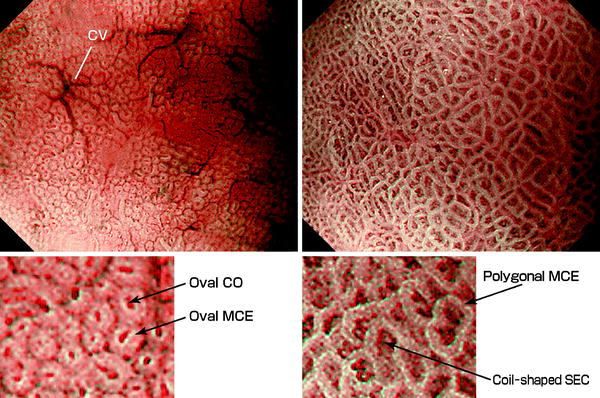
Fig. 11.1
Case 1: M-NBI findings of the normal fundic glandular epithelium (left) and normal gastric pyloric glandular epithelium (right) (GIF-Q240Z, maximal magnifying ratio)
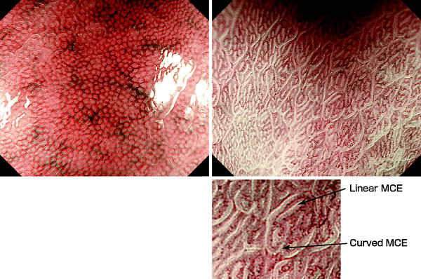
Fig. 11.2
Case 2: M-NBI findings of the normal fundic glandular epithelium (left) and nor mal pyloric glandular epithelium (right) (GIF-H260Z, maximal magnifying ratio)
11.2.1.1 V: Microvascular Architecture
Anatomically, the microvascular architecture is formed by the capillaries and collecting venules (CVs). The morphology of each capillary is that of a dark brown polygonal closed loop. These loops anastomose repeatedly with each other, forming a regular honeycomb-like subepithelial capillary network (SECN) pattern. In the normal fundic glandular epithelium, almost 100 % of the time, regularly arranged cyan-colored collecting venules (CVs) can be identified. We can see the regular SECN draining into CVs in the superficial mucosal layers [1, 2].
11.2.1.2 S: Microsurface Structure
Anatomically, the mucosal surface is made up of the marginal crypt epithelium (MCE), crypt openings (COs), and the intervening part (IP) in between. The epithelial morphology is visualized as semitransparent white belt-like structures, showing a circular or oval shape. In the center we see brown oval COs, surrounded by MCE.
11.2.1.3 Combined Morphology of V and S
The VS (relationship between the microvascular architecture and microsurface structure) is an epithelium within vessel pattern, with oval areas of epithelium seen within polygonal subepithelial capillaries (SECs). For convenience’s sake, we refer to this pattern of oval epithelial areas inside blood vessel closed loops as “VS of gastric body type.”
11.2.2 Gastric Antrum (Pyloric Type Glandular Epithelium) (Figs. 11.1 and 11.2) [1, 2]
11.2.2.1 V: Microvascular Architecture
Anatomically, the microvascular architecture is formed by the capillaries and CVs, but the latter are rarely observed from the mucosal surface [3, 4]. The morphology of each capillary is that of dark brown coil-shaped open loops. Although it has been shown anatomically that these loops anastomose repeatedly with each other under the epithelial surface, subepithelial anastomoses are not often visualized. Only rarely do the capillaries anastomose together to form a subepithelial reticular pattern.
11.2.2.2 S: Microsurface Structure
Anatomically the mucosal surface is made up of the MCE and the IPs surrounded by the MCE. As described in earlier chapters, we rarely observe dark brown COs in the antral mucosa. The MCE morphology is usually polygonal but may be curved or linear.
11.2.2.3 Combined Morphology of V and S
The VS is a vessel within epithelium pattern, with coil-shaped SECs within polygonal MCE. For convenience’s sake, we refer to this pattern as “VS of gastric antral type.”
11.3 Early Gastric Cancer
11.3.1 Summary
Advantages and problems using NBI with ME [5].
 Basic Principles for the Interpretation of Magnified Endoscopic (ME) Findings: Vessels (V) Plus Surface (S) Classification System
The Proposed Vessels Plus Surface (VS) Classification System: Principles for Interpretation of Magnifying Endoscopy with Narrow-Band Imaging (M-NBI) Findings
Basic Principles for the Interpretation of Magnified Endoscopic (ME) Findings: Vessels (V) Plus Surface (S) Classification System
The Proposed Vessels Plus Surface (VS) Classification System: Principles for Interpretation of Magnifying Endoscopy with Narrow-Band Imaging (M-NBI) Findings
 Microanatomies as Visualized Using Magnifying Endoscopy with Narrow Band Imaging in the Stomach: Which Microanatomical Structures Can We Visualize in the Glandular Epithelium Using Narrow Band Imaging, and How Is This Achieved?
 Light Blue Crests (LBCs) and White Opaque Substance (WOS)
 Magnifying Endoscopy (ME) of the Stomach Targeting the Microvascular Architecture
 Analysis and Interpretation of Magnifying Endoscopy with Narrow Band Imaging (M-NBI) Findings in Gastric Epithelial Tumors (Early Gastric Cancer and Adenoma) Stratified for Paris Classification of Macroscopic Appearance


Advantages
< div class='tao-gold-member'>
Only gold members can continue reading. Log In or Register to continue
Related posts:
Basic Principles for the Interpretation of Magnified Endoscopic (ME) Findings: Vessels (V) Plus Surface (S) Classification System
The Proposed Vessels Plus Surface (VS) Classification System: Principles for Interpretation of Magnifying Endoscopy with Narrow-Band Imaging (M-NBI) Findings
Microanatomies as Visualized Using Magnifying Endoscopy with Narrow Band Imaging in the Stomach: Which Microanatomical Structures Can We Visualize in the Glandular Epithelium Using Narrow Band Imaging, and How Is This Achieved?
Light Blue Crests (LBCs) and White Opaque Substance (WOS)
Magnifying Endoscopy (ME) of the Stomach Targeting the Microvascular Architecture
Analysis and Interpretation of Magnifying Endoscopy with Narrow Band Imaging (M-NBI) Findings in Gastric Epithelial Tumors (Early Gastric Cancer and Adenoma) Stratified for Paris Classification of Macroscopic Appearance
Stay updated, free articles. Join our Telegram channel
Full access? Get Clinical Tree
