(1)
Department of Endoscopy, Fukuoka University Chikushi Hospital, Fukuoka, Chikushino, Japan
Summary
Gastric body mucosal patterns identified using magnifying endoscopy (ME)
Type 1
V: Regular honeycomb-like SECN pattern with regular CV pattern present
S: Regular oval crypt opening pattern
Type 2
V: Regular honeycomb-like SECN pattern but regular CV pattern absent
S: Regular oval/tubular crypt opening pattern
Type 3
V: Loss of regular honeycomb-like SECN pattern and regular CV pattern absent
S: Regular but enlarged white oval/tubular crypt opening pattern
Type 4
V: Loss of regular honeycomb-like SECN pattern with slightly irregular CV pattern present
S: Flat or non-structured, with microsurface pattern absent
Keywords
Chronic gastritisClassificationGastric bodyMagnifying endoscopyStomachExplanation
The gastric mucosal ME findings of chronic gastritis associated with Helicobacter pylori (H. pylori) infection encompass wide variations in microvascular architecture (V) and microsurface structure (S) and have yet to be fully elucidated. Some ME findings in the gastric body suitable for clinical application have been published, however, so in this chapter I will present these and save discussion of light blue crests for Chap. 10.
There have been reports of the usefulness of ME of the gastric body mucosa in assessing the degree of severity of chronic gastritis and whether H. pylori infection is present [1–3]. The above categories were developed for an investigation of the reproducibility of ME findings of H. pylori infection and atrophy in the gastric body by myself and some colleagues in the UK in 2005. These were based on earlier studies by Yagi [2] and Nakagawa [1], and I strongly recommend that you refer to their original papers.
Putting it simply, normal gastric body mucosa free of H. pylori infection is always characterized by V, regular honeycomb-like SECN pattern with regular CV pattern present, plus S, regular oval crypt opening pattern (Fig. 5.1). H. pylori infection causes loss of the CVs (Figs. 5.2 and 5.3), and with atrophic gastritis (Fig. 5.4) we can again see the CVs, but the morphology and distribution of the CVs becomes irregular.
 Basic Principles for the Interpretation of Magnified Endoscopic (ME) Findings: Vessels (V) Plus Surface (S) Classification System
The Proposed Vessels Plus Surface (VS) Classification System: Principles for Interpretation of Magnifying Endoscopy with Narrow-Band Imaging (M-NBI) Findings
Basic Principles for the Interpretation of Magnified Endoscopic (ME) Findings: Vessels (V) Plus Surface (S) Classification System
The Proposed Vessels Plus Surface (VS) Classification System: Principles for Interpretation of Magnifying Endoscopy with Narrow-Band Imaging (M-NBI) Findings
 Microanatomies as Visualized Using Magnifying Endoscopy with Narrow Band Imaging in the Stomach: Which Microanatomical Structures Can We Visualize in the Glandular Epithelium Using Narrow Band Imaging, and How Is This Achieved?
 Light Blue Crests (LBCs) and White Opaque Substance (WOS)
 Magnifying Endoscopy (ME) of the Stomach Targeting the Microvascular Architecture
 Analysis and Interpretation of Magnifying Endoscopy with Narrow Band Imaging (M-NBI) Findings in Gastric Epithelial Tumors (Early Gastric Cancer and Adenoma) Stratified for Paris Classification of Macroscopic Appearance


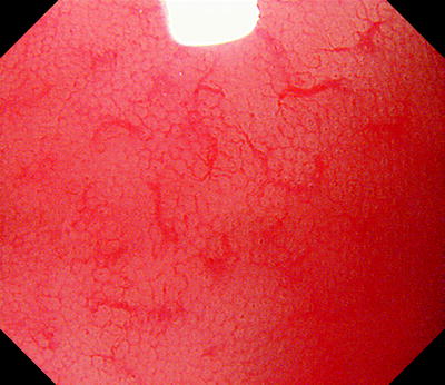
Fig. 5.1
Type 1: in our study, this pattern identified H. pylori negative normal gastric mucosa with 100 % positive predictive value. This corresponds to the R pattern of Nakagawa et al.
< div class='tao-gold-member'>
Only gold members can continue reading. Log In or Register to continue
Related posts:
Basic Principles for the Interpretation of Magnified Endoscopic (ME) Findings: Vessels (V) Plus Surface (S) Classification System
The Proposed Vessels Plus Surface (VS) Classification System: Principles for Interpretation of Magnifying Endoscopy with Narrow-Band Imaging (M-NBI) Findings
Microanatomies as Visualized Using Magnifying Endoscopy with Narrow Band Imaging in the Stomach: Which Microanatomical Structures Can We Visualize in the Glandular Epithelium Using Narrow Band Imaging, and How Is This Achieved?
Light Blue Crests (LBCs) and White Opaque Substance (WOS)
Magnifying Endoscopy (ME) of the Stomach Targeting the Microvascular Architecture
Analysis and Interpretation of Magnifying Endoscopy with Narrow Band Imaging (M-NBI) Findings in Gastric Epithelial Tumors (Early Gastric Cancer and Adenoma) Stratified for Paris Classification of Macroscopic Appearance
Stay updated, free articles. Join our Telegram channel
Full access? Get Clinical Tree
