(1)
Department of Vascular Surgery, Evangelisches Krankenhaus Königin Elisabeth Herzberge, Berlin, Germany
Apart from meeting the diverse biomechanical standards, a catheter should also guarantee a blood flow of at least 250–300 mL/min.
There is a definite distinction between temporary and permanent catheters.
2.1 Temporary Catheters
Indication
Access for a limited period of time (3–4 weeks at most) until a permanent vascular access or access for peritoneal dialysis can be used, or until dialysis is no longer necessary.
Location
Every catheter may cause venous thrombosis. The risk for thrombosis depends on the:
Anatomical position of the vein
Period of time for which the catheter has been used
Length of the intraluminal segment of the catheter
Diameter of the catheter
Thrombogenicity of the material
Possible coagulation disorders
Bacterial colonisation of the catheter
Intimal irritation and the risk of thrombosis are much lower in the internal jugular vein than in the subclavian or femoral veins. Criteria for the selection of a suitable vein include the:
Patency of the vein
Local conditions (e.g., infections, scar tissue)
Risk of thrombosis
Clinical sequelae of a previous thrombosis (e.g., AV access of the ipsilateral arm no longer feasible after thrombotic occlusion of the subclavian vein)
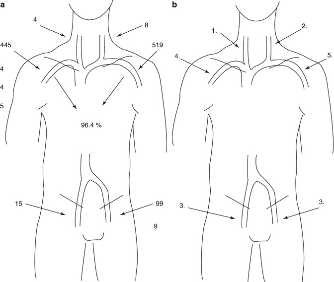
The analysis of the last 1,000 AV access procedures in our patients has shown that in 96.4 % the subclavian veins were used as the venous runoff (Fig. 2.1a). Therefore the protection of these veins is quite important. The following recommendations regarding the sequence for temporary catheter placement are based on this experience (Fig. 2.1b):


Fig. 2.1
(a ) Total numbers of patients treated by that specific way out of 1,000. (b) Recommended sequence for placement of central venous catheters
1.
Right internal jugular vein (shortest intraluminal segment of catheter)
2.
Left internal jugular vein (if right internal jugular vein not available)
3.
Common femoral vein (to preserve the subclavian veins if internal jugular veins not suited)
4.
Right subclavian vein (if jugular and femoral veins not available)
5.
Left subclavian vein (if jugular, femoral, and right subclavian veins not available)
A thrombosis of the left subclavian vein often coincides with a thrombosis of the left brachiocephalic vein. On the right side this co-occurrence is extremely rare.
Types of Catheters
The higher mean flow (250–300 mL/min) of double lumen catheters (external diameter 11–12 Ch) constitutes an advantage over single lumen catheters (external diameter 8 Ch) with flow volumes of around 200 mL/min. There are also high flow catheters with external diameters of 13 Ch, which grant even higher flows (around 400 mL/min). Polyurethane and silicone are common catheter materials.
Stay updated, free articles. Join our Telegram channel

Full access? Get Clinical Tree


