, Mark McAlindon6 , Dan Carter10 , Rami Eliakim7 , Anastasios Koulaouzidis8 , Sarah Douglas8 , Wenbin Zou1 , Zhizheng Ge9 and Zhaoshen Li1
(1)
Department of Gastroenterology, Changhai Hospital, Second Military Medical University, 168 Changhai Road, Yangpu District, Shanghai, 200433, China
(2)
Department of Gastroenterology, The First People’s Hospital of Yunnan Province, Kunming, China
(3)
Department of Gastroenterology, General Hospital Affiliated to Tianjin Medical University, Tianjin, China
(4)
Department of Gastroenterology, The Third Affiliated Hospital of Southern Medical University, Guangzhou, 510630, China
(5)
Department of Gastroenterology, Nanfang Hospital, Southern Medical University, Guangzhou, 510515, China
(6)
Gastroenterology, Royal Hallamshire Hospital, Sheffield Teaching Hospitals NHS Trust, Glossop Road, Sheffield, S10 2JF, UK
(7)
Department of Gastroenterology, Sheba Medical Center, Sackler School of Medicine, Tel-Aviv University, Tel-Aviv, Israel
(8)
The Royal Infirmary of Edinburgh, Centre for Liver and Digestive Disorders, 51 Little France Crescent, Edinburgh, EH16 4SA, UK
(9)
Department of Gastroenterology and Hepatology, Digestive Endoscopy Centre, Renji Hospital, School of Medicine, Shanghai Jiao Tong University, Shanghai, China
(10)
Department of Gastroenterology, Chaim Sheba Medical Center, 2nd Sheba Road, 52621 Ramat-Gan, Israel
10.1 Small Bowel Capsule Endoscopy I
Case 1. Cavernous hemangioma in jejunum.
A 74-year-old Chinese woman with recurrent melena for 7 years. Her hemoglobin level was 90 g/L (Fig. 10.1).
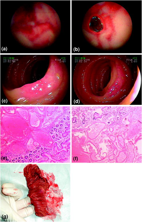

Fig. 10.1
a, b SBCE revealed the hemorrhagic lesions in the upper of jejunum; c, d hemangiomas were suspected under double-balloon enteroscopy in the jejunum; e, f pathological findings confirmed the diagnosis of cavernous hemangioma of jejunum; and g surgical specimens
Case 2. Meckel’s diverticulum .
A 21-year-old Chinese woman was admitted for recurrent melena. Gastroscopy and colonoscopy revealed no hemorrhagic lesion. Her hemoglobin level was 115 g/L (Fig. 10.2).
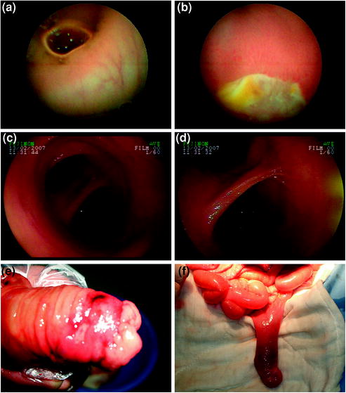

Fig. 10.2
a, b SBCE found ileal ulcers; c, d double-balloon enteroscopy confirmed Meckel’s diverticulum ; and e, f surgical specimen
Case 3. Rupture of ileum venous aneurysm .
A 49-year-old Chinese woman presented with recurrent melena and hematochezia for a month. Her hemoglobin level was 69 g/L. Colonoscopy found only intraluminal blood with no abnormal mucosa (Fig. 10.3).
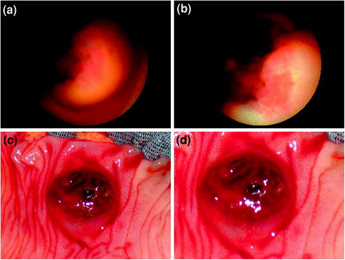

Fig. 10.3
a, b SBCE found elevated lesions bleeding in the upper ileum, suspecting hemangioma or stromal tumor s and c, d surgery confirmed venous aneurysm bleeding in the upper ileum
Case 4. Mucosa-associated B-cell lymphoma in the small intestine.
A 51-year-old Chinese woman had melena twice. She was admitted for hematochezia (Fig 10.4).
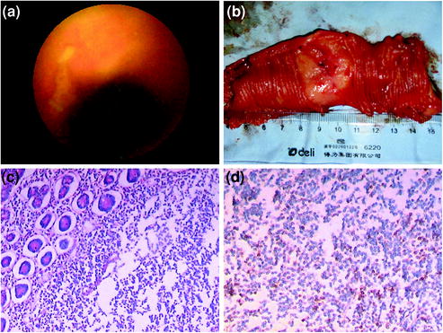

Fig. 10.4
a SBCE found ileal ulcers; b the resected tumor was located in the jejunum–ileum junction; c pathological findings were that lymphoid tissue diffusely infiltrated the whole intestinal wall, suspecting lymphoma; and d immunohistochemical stains of resected tumor were positive for CD3, CD20, CD45RO, CD45RA, CD5, CyclinD1, and bcl-2
Case 5. Jejunum vascular malformation .
A 15-year-old Chinese boy had melena accompanying with syncope for a week. He was admitted for a sudden severe melena with hemoglobin of 70 g/L, which led to shock (Fig. 10.5).
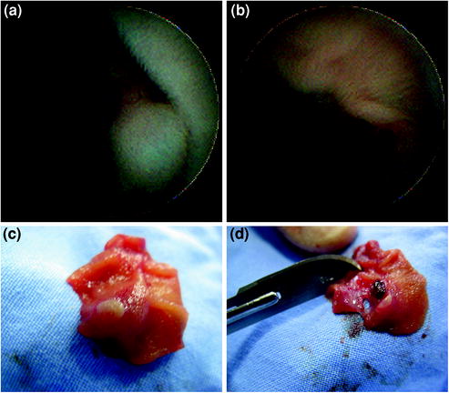

Fig. 10.5
a, b SBCE showed a vascular lesion with blood clots in the mid–upper jejunum and c, d surgical specimen confirmed the diagnosis of vascular malformation
Case 6. Hookworms in duodenum.
A 45-year-old Chinese man had melena for a week (Fig. 10.6).
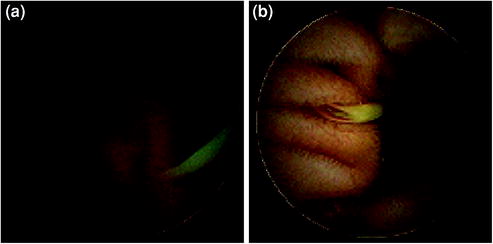

Fig. 10.6
a, b SBCE found hookworms in the duodenum
Case 7. Taeniasis in small intestine.
A 58-year-old Chinese man presented with recurrent abdominal pain for three years (Fig. 10.7).
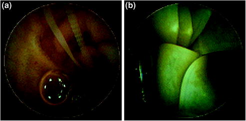

Fig. 10.7
a, b SBCE found tapeworm in the small intestine, diagnosing Taeniasis
Case 8. Stromal tumor s in mid-ileum.
A 57-year-old Chinese woman had melena with hemoglobin of 70 g/L for a week (Fig. 10.8).
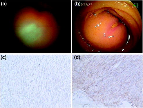

Fig. 10.8


a SBCE showed an elevated lesion in mid-ileum; b Double-balloon enterography findings; and c, d pathological and Immunohistochemical stains of resected tumor confirmed the diagnosis of stromal tumor in mid-ileum
Stay updated, free articles. Join our Telegram channel

Full access? Get Clinical Tree


