| 2 | Basic Examination Technique and Colonoscopy Workstation |
Learning Examination Technique
Learning colonoscopy technique requires motivation, manual dexterity, concentration, and patience on the part of the trainee. Already practiced in the technique of endoscopically examining the upper digestive tract, the beginner will learn about similarities and differences related to using a gastroscope vs. using a colonoscope.
 Instrument Features
All endoscopes can be divided into three sections: the insertion tube, which is advanced in the patient, the instrument control head, where the physician can maneuver the endoscope tip and has access to the water/air supply and the instrument channel and, finally, the universal cord and plug, which connect the instrument to the supply unit (Fig. 2.1).
Insertion tube. The insertion tube of the video colonoscope consists of a ca. 130-cm-long tube containing optical fibers, digital wires, air and water nozzles, the instrument channel, and Bow-den cables for better mobility.
Located at the tip of the endoscope are the lens and video chip, which produces the image (Fig. 2.2).
The last 15 cm of the insertion tube are especially flexible and can bend in all four directions, allowing better maneuvering in the gastrointestinal tract. The degree of flexion of the colonoscope is 180° up/down and 160° to the right/left. Compared with the colonoscope, the gastroscope can be moved to a greater degree upward, but to a lesser degree in all other directions (Figs. 2.3, 2.4, Tab. 2.1).
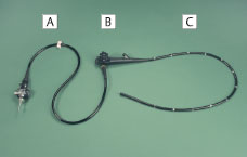
Fig. 2.1 Main parts of the colonoscope. A Universal cord and plug, B Instrument control head, C Insertion tube (courtesy of Mr. Wirth, photo archive, Augsburg Clinic).
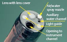
Fig. 2.2 Tip of colonoscope (courtesy of Mr. Wirth, photo archive, Augsburg Clinic).
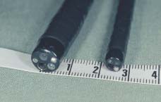
Fig. 2.4 Comparing the outer diameters of the distal end of a colonoscope (left) and a gastroscope (right) (courtesy of Mr. Wirth, photo archive, Augsburg Clinic).
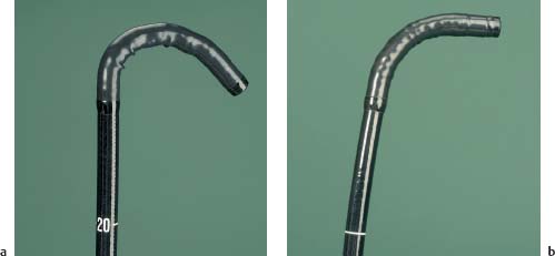
Fig. 2.3 Maximum angling downward.
a Colonoscope.
b Gastroscope.
(Courtesy of Mr. Wirth, photo archive, Augsburg Clinic.)
Standard video endoscopes | Colonoscope | Gastroscope |
|---|---|---|
Maximum angling | Up 180° | Up 210° |
Down 180° | Down 90° | |
Right/left 160° | Right/left 100° | |
Outer diameter, distal end | 12.8 mm | 10.2 mm |
Length of insertion tube | 103 cm | 103 cm |
Angle | 140° | 130° |
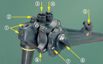
Fig. 2.5 Control head of a video colonoscope:
1 Function buttons, e.g., video recorder remote control
2 Freeze button
3 Suction button
4 Air/water button
5 Instrument channel
6 Locking device
7 Angling wheel (right/left)
8 Angling wheel (up/down)
(Courtesy of Mr. Wirth, photo archive, Augsburg Clinic.)
The sigmoidoscope measures only 60 cm in total length. Because of its high degree of maneuverability, it is sometimes used in patients where the indications for examination are limited to the sigmoid colon and rectum.
Different colonoscope models can vary in length, outer diameter, and width of the instrument channel.
Instrument control head. The functions necessary for maneuvering the tip the endoscope, for suction, cleansing, and air insufflation are all located on the control head. (Fig. 2.5).
The opening to the instrument channel is somewhat below the air/water cylinder, but before the air/water channel merges with the suction channel. The diameter of the inside of the instrument channel is between 2.8 mm and 3.7 mm, allowing the insertion of endoscopic accessories such as biopsy forceps or polypectomy snares (Fig. 2.6).
Video endoscopes also have remote control buttons that, according to model, may have various functions. These buttons can generally be used for freeze frames, video recording, printing, and adjusting illumination intensity (peak and average). Newer generations are equipped with so-called big chips that allow the projection of a high-resolution screen-size image onto a video monitor. The image can be digitally enhanced using modern image processing technology (e.g., Olympus CV-160) for structure enhancement, variable by several levels during and even after the examination (see Chapter 3).
Universal cord. The universal cord connects the endoscope to the light source, air supply, water supply, suction pump, and video processor. The video processor transmits the image to the monitor screen on the video tower (Figs. 2.7–2.10).
 Operating the Endoscope
Before every examination, the suction, cleansing, and air insufflation functions on the endoscope must be checked and the “white balance” set on the video processor. The physician’s left hand holds the control head of the endoscope while the right hand moves the insertion tube or controls fine adjustment of the outer angling wheel on the control head (Fig. 2.11).
Suction and cleansing. The index finger of the left hand can be used to depress the suction button while the middle finger can either press the air/water button lightly for insufflation or more firmly to activate the washing system (Fig. 2.12).
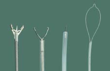
Fig. 2.6 Colonoscopy accessories (from left to right): biopsy forceps, clip applicator, injection needle, and polypectomy snare (courtesy of Mr. Wirth, photo archive, Augsburg Clinic).
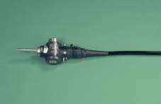
Fig. 2.7 Universal plug on an endoscope (courtesy of Mr. Wirth, photo archive, Augsburg Clinic).
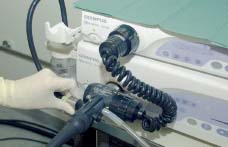
Fig. 2.8 Plugging the universal cord into the processing unit (courtesy of Mr. Wirth, photo archive, Augsburg Clinic).
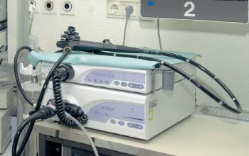
Fig. 2.9 Video processor (above) and light source (below) (courtesy of Mr. Wirth, photo archive, Augsburg Clinic).
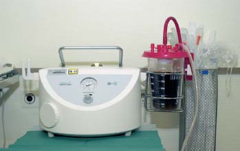
Fig. 2.10 Suction pump (courtesy of Mr. Wirth, photo archive, Augsburg Clinic).
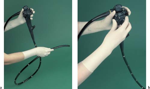
Fig. 2.11 Operating the instrument.
a The examiner’s right hand guides the tube or
b Fine adjustment of the endoscope tip by moving the angling wheel.
Stay updated, free articles. Join our Telegram channel

Full access? Get Clinical Tree


