Fig. 7.1
Balloon-assisted enteroscopy. (a) Double-balloon enteroscope. (b) Single-balloon enteroscope. The major similitude is the overtube with the balloon (red circle). In double-balloon enteroscopy there is an additional balloon on top of the scope (yellow oval). Note balloon insufflators for Fujinon (c) and Olympus systems (d), respectively
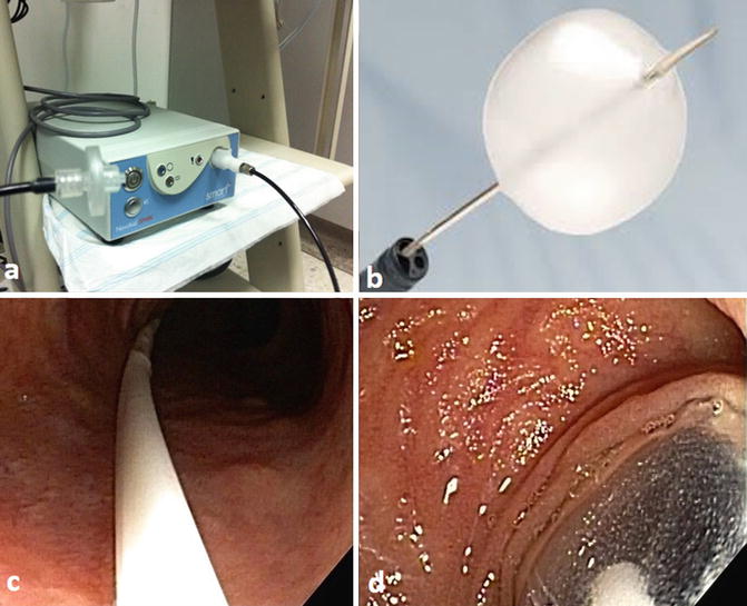
Fig. 7.2
Balloon-guided enteroscopy (BGE). The BGE device is attached to the scope. (a) Inflation device. (b) Advancing balloon. (c) Endoscopic view of advancing catheter. (d) Endoscopic view of advancing balloon
Indications
The indications for BAE have increased significantly since its introduction in 2004. Currently, BAE techniques are not only used to perform deep enteroscopy, but the equipment can also be used for endoscopic retrograde cholangiopancreatography (ERCP), incomplete colonoscopies, or exploring the gastrointestinal tract of patients with surgically deranged anatomy [16–28] (Fig. 7.3). Table 7.1 lists indications for BAE. Furthermore, BAE has become an important tool to deliver various types of endoscopic therapies during deep enteroscopy, but also for many other gastrointestinal problems, such as insertion of self-expanding metal stents into the small bowel or into the stomach in patients with deranged anatomy [22–26] (Table 7.1, Figs. 7.4 and 7.5).
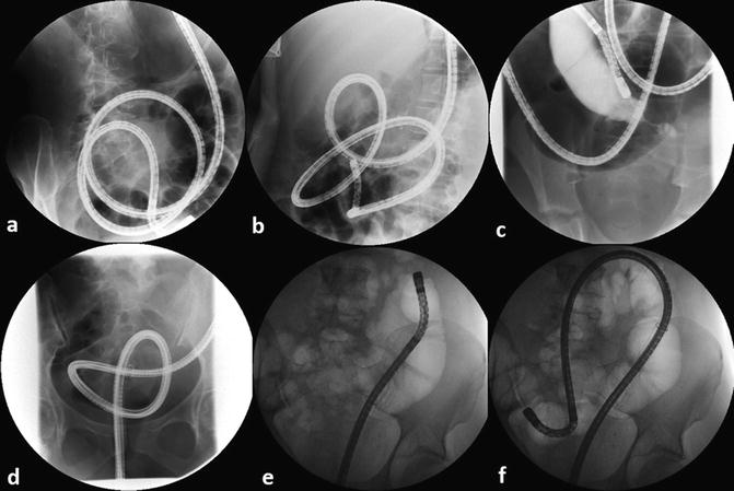
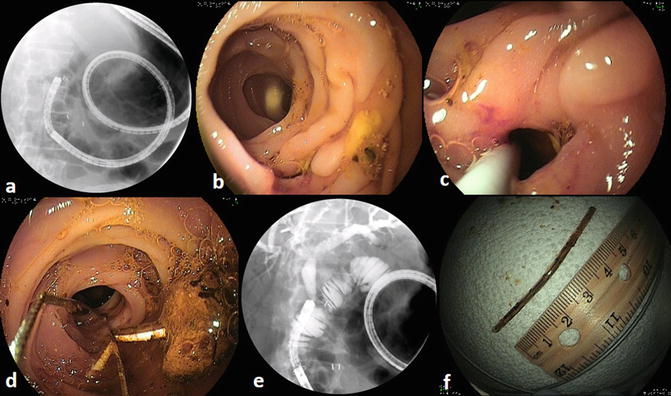
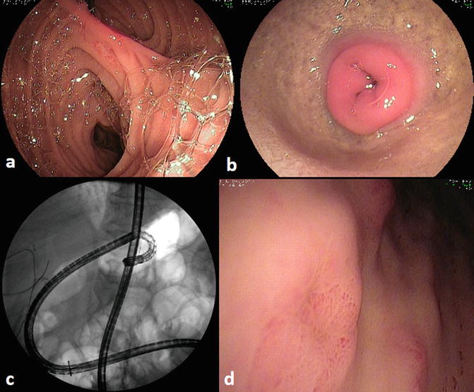

Fig. 7.3
Fluoroscopy during balloon-assisted enteroscopy (BAE). The images show (a) antegrade (oral) BAE, (b) looping of bowel due to adhesions during an oral BAE, (c) use of contrast to delineate a stricture in a patient with Crohn’s disease, (d) “pretzel” configuration of the scope in a patient with previously failed colonoscopy, (e) retrograde (anal) BAE, and (f) passing of a long and floppy splenic flexure during anal BAE
Table 7.1
Indications and potential therapeutic interventions using the balloon-assisted enteroscopy
Small bowel bleeding Hemostasis • Argon plasma coagulation • Injection of diluted epinephrine (1:100,000) • Sclerotherapy (cyanoacrylate injection, Dermabond®, Histoacryl®) • Placement of clips (standard hemoclips or over-the-scope clips) |
Crohn’s disease Balloon dilation of strictures Retrieval of retained small bowel capsule |
NSAID enteropathy Balloon dilation of strictures |
Malabsorption syndromes |
Celiac disease (surveillance) |
Polyposis syndromes (surveillance) |
Polypectomy |
Endoscopic mucosal resection |
Tumors (adenocarcinoma, gastrointestinal stromal tumors, search for primary neuroendocrine tumors) |
Submucosal injection with India ink |
Removal of foreign bodies (e.g., capsule endoscope, pins, dentures, needles, coins) |
Colonoscopy Complete a previously failed colonoscopy Stent placement |
Percutaneous endoscopic jejunostomy in normal and altered bowel anatomy (gastric bypass, Roux-en-Y) |
Biliary interventions ERCP • Cholangiogram • Pancreatogram • Sphincterotomy • Precut sphincterotomy • Papillectomy • Balloon dilation • Stone removal • Stent placement/removal (plastic and metal stents) • Removal of dislocated metal stent |

Fig. 7.4
BAE-ERCP in a patient with Whipple operation and hepaticojejunostomy with Roux-en-Y anastomosis. (a) Scope in biliodigestive limb. (b) The hepaticojejunostomy is often difficult to find. Here it is located at the 6 o’clock position. (c) Cannulation of the hepaticojejunostomy. (d) Removal of sludge and an inwardly migrated plastic stent with a basket. (e) Cholangiography. (f) Removed stent

Fig. 7.5
BAE in a patient with Roux-en-Y gastric bypass. (a) Recognition of the afferent loop is facilitated by the presence of many small and large yellowish air bubbles, which result from the presence of bile. (b) Pylorus viewed from the “back.” (c) Retroflexion of the enteroscope inside of the excluded stomach. The retroflexion was performed to visualize the antrum. (d) Multiple ulcers and erosions were seen
Equipment
We will separately describe the equipment and technique used for overtube-based BAE and through-the-scope balloon-guided enteroscopy.
Overtube-Assisted BAE
Currently there are two types of balloon enteroscopes: DBE (Fujifilm, Japan) and SBE (Olympus Optical Co, Ltd, Tokyo, Japan) [1–8, 13, 29, 30] (Fig. 7.1). The enteroscopes used to perform DBE come in various lengths and diameters (Table 7.2). This variety allows one to choose the scope for different conditions. The diagnostic or small-diameter enteroscope is potentially useful for narrowed luminal diameter, significant adhesions, and/or performing incomplete colonoscopy [13, 30]. However, the small working channel of this enteroscope limits the therapeutic capabilities, as there are few accessories that can be easily advanced through this 2.2 mm channel. For this reason, most experts prefer the “therapeutic” enteroscope, which has a larger diameter and wider working channel (2.8 mm) [29, 30] (Table 7.2, Video 7.1). The single-balloon enteroscope (SIF Q260) has a 200 cm working length, a 9.2 mm outer diameter, and a 2.8 mm working channel [5–8]. This enteroscope from Olympus and the “diagnostic” and “therapeutic” enteroscopes from Fujifilm have the same length, in contrast to the short enteroscopes from Fujifilm (Table 7.2) [1–8, 13, 24]. The advantage of the shorter enteroscope is its utility for ERCP [27, 28, 30]. To simplify the definitions, we call the long enteroscopes “standard” length scopes and the shorter scopes “short enteroscopes.” In addition, the reader should know that the DBE scopes could also be used to perform SBE [29]. In this instance, no balloon is attached to the endoscope itself.
Table 7.2
Enteroscopy device specifications
Specifications | |||||
|---|---|---|---|---|---|
Scopes | Scope working length (cm) | Scope diameter (mm) | Channel diameter (mm) | Overtube length (cm) | Overtube outer diameter (mm) |
SBE (Olympus, SIF-Q180) | 200 | 9.2 | 2.8 | 140 | 13.2 |
DBE (Fujinon, EN-450PS/20) | 200 | 8.5 | 2.2 | 145 | 12.2 |
DBE (Fujinon, EN-450T5) | 200 | 9.4 | 2.8 | 145 | 13.2 |
DBE (Fujinon, EC-450BI5) | 152 | 9.4 | 2.8 | 105 | 13.2 |
Pentax VSB-3,430 K | 220 | 11.6 | 3.8 | ||
SE Spirus medical, discovery SB | a | a | a | 118 | 16 |
Both standard DBE enteroscopes and their respective overtubes have the same length (enteroscope 200 cm, overtube 1,450 mm), but the external diameters of both the endoscope and the overtube are different. The diagnostic DBE (EN-450P5, Fujinon, Saitama, Japan) has an external diameter of 8.5 mm and the overtube has a diameter of 12.2 mm, whereas the therapeutic DBE (EN-450T5) has an external diameter of 9.4 mm and the overtube has a diameter of 13.2 mm [13, 24, 30]. The diameter of the working channel of the therapeutic DBE is 2.8 mm whereas the diagnostic one is 2.2 mm wide [13, 24, 29, 30]. Thus, careful attention should be paid when choosing accessories, as these must be long enough to exit the scope and also have an external diameter that permits their insertion and pushing through the respective working channels. In our endoscopy unit we keep a list of the sizes, diameters, lengths, and working channel capabilities of all the endoscopes we use. These tables are posted on the wall of each room and serve as a quick reference. The lengths and sizes of the overtubes are also specified in Table 7.2.
Technique of Balloon-Assisted Enteroscopy
We prefer to use general anesthesia when performing oral (antegrade) BAE. When using general anesthesia we prefer to place the patient on their back (i.e., supine). This position will also allow for easy external manual compression when this is needed to improve advancement of the scope. When conscious sedation is utilized, left-lateral decubitus positions are mandatory, so that the patient’s secretions can be managed properly and thus aspiration avoided [13]. We use moderate sedation for the majority of our patients undergoing anal (retrograde) BAE.
Both techniques of SBE and DBE, similar to overtube-assisted push enteroscopy, use the principle of the push-and-pull technique using the overtube: The endoscope is first inserted (pushed) into the small bowel, then the overtube is advanced toward the tip of the endoscope, and then both endoscope and overtube are pulled back; hence the name “push and pull” (Fig. 7.6) [13]. This standard approach to BAE requires two people, the operator and an assistant. The assistant holding the overtube plays a crucial role, as a stable overtube will positively influence the depth of insertion and the maneuverability of the scope [13]. When using SBE and DBE, the major factor governing the depth of insertion of the enteroscope is the presence of the balloon on the distal end of the flexible overtube. The overtube stabilizes the intestine, preventing it from bending or looping [13, 30]. Although, the overtube stabilizes the intestine, preventing it from bending or looping, the key action of the inflated overtube balloon is to shorten the small bowel proximally (Fig. 7.6a). The inflated balloon prevents the intestine from sliding forward while the scope is being pushed or advanced forward (Fig. 7.6b) [13, 30]. This allows the endoscopist to firmly push on the enteroscope, effectively transmitting forces to the distal end of the endoscope and hence permitting the advancement of the enteroscope deeper into the small bowel without looping or stretching of the proximal intestine [1–8, 10, 13, 30]. The key steps and aspects of BAE are listed in Table 7.3 and shown in Fig. 7.6 and Video 7.2.
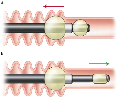

Fig. 7.6


Key elements of double-balloon enteroscopy. (a) Both the overtube and scope are pulled back (red arrow) to straighten the bowel and retract the proximal bowel on top of the overtube. (b) The balloon of the overtube is kept inflated (keeping the retracted bowel in place) and the balloon of the scope is deflated. The inflated balloon prevents the intestine to slide forward, keeping it “shortened” while the scope is being pushed or advanced forward (green arrow shows direction of scope)
Stay updated, free articles. Join our Telegram channel

Full access? Get Clinical Tree


