Fig. 2.1
Overview of the pelvic floor muscles and anal sphincters seen from below
The pubococcygeus originates from the posterior inferior aspect of the pubis and the inner anterior surface of the obturator fascia, including a portion of the arcus tendineus of the levator ani. Its anterior fibers insert medially into the central tendon of the perineum (hiatal ligament), where they fuse with the musculature of the prostate, vagina, and perineal body to form the levator prostate, pubovaginalis, and pubourethralis muscles. Some of these intermediate fibers also travel caudally along the intersphincteric plane and contribute to the conjoined longitudinal coat of the anal canal. The muscular fibers of the pubococcygeus merge posteriorly into the broad fibrous band that inserts into the anococcygeal ligament, anterior sacrococcygeal ligament, and coccyx.
The iliococcygeus arises from the arcus tendineus of the fascia of the internal obturator muscle posterior and caudal to the origin of the pubococcygeus. The fibers run posteromedially, where they merge and insert into the anococcygeal ligament and the last two segments of sacrum.
The puborectalis is the most caudal and controversial component of the levator ani complex. It arises from the posterior aspects of the body of the pubis, the inferior pubic ramis, the superior fascia of the urogenital diaphragm, and the adjacent obturator internus fascia and loops around the rectum to form a strong U-shaped sling. Medial fibers of the puborectalis fan out and insert into the central tendon of the perineum where they intermingle with fibers from the pubococcygeus and contribute to the conjoined longitudinal muscle of the anal canal. The puborectalis sling, together with the upper borders of the internal and external sphincters, forms the anorectal ring, which delineates the surgical anal canal from the rectum.
Rectum and Anal Canal
The rectum begins at the level of the third sacral vertebra and in general follows along the curvature of the sacrum and coccyx for its entire length of 12–15 cm (Fig. 2.2). In addition, it has three lateral curves; the upper and lower ones are convex to the right, the middle one convex to the left. The inner aspects of these three transverse infoldings correspond to the rectal valves of Houston. At the rectosigmoid junction, the fibers of the taenia spread out to form the longitudinal muscle layer of the rectum, which surrounds the inner circular muscle layer. The outer muscle coat is slightly thicker on the anterior and posterior rectal walls than on its lateral surfaces, contributing to the formation of three lateral curves.


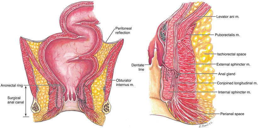
Fig. 2.2
Coronal view of the anal canal, lower rectum, and surrounding spaces. The enlarged view on the right highlights the architecture of the sphincter muscles and anal glands
The upper surface of the rectum is covered by the peritoneum on its anterior and lateral surfaces. The middle third is covered on its anterior surface only, and the lower third is entirely below the peritoneal reflection. The middle valve of Houston is approximately at the level of the anterior peritoneal reflection.
The posterior wall of the rectum is covered with a thick layer of pelvic fascia (fascia propria). A strong sheet of fascia (Waldeyer’s or rectosacral fascia) arises from the fourth sacral segment, tracks forward and downward, and attaches to the fascia propria on the posterior surface of the rectal wall at the anorectal junction. The lower portion of the rectum is supported on each side by reflections of endopelvic fascia known as the lateral stalks of the rectum. The anterior extraperitoneal surface of the rectum is covered by Denonvilliers’ visceral pelvic fascia which extends to the urogenital diaphragm running between the rectum and prostate or vagina.
As the rectum passes through the pelvic diaphragm at the level of the anorectal ring, it changes its name, shape, and direction. The anal canal begins at this point and extends for approximately 4 cm to the anal verge (surgical anal canal). The circular lumen of the rectum flattens into an anteroposterior slit because of its attachment to the perineal body and coccyx along with the medial pressure exerted by the ischiorectal fat pads. The puborectalis sling in its normal contracted state angulates the anorectal junction forward to create an 80° bend, the perineal flexure, which may assist the external sphincter mechanism in maintaining fecal continence.
The internal sphincter is a continuation of the involuntary layer of circular smooth muscle of the rectum that begins at the level of the anorectal ring. As it proceeds distally, it becomes appreciably thicker, and its rounded lower margin can usually be palpated about 1–2 cm below the dentate line. The internal sphincter, like the puborectalis, is tonically contracted “at rest”.
The conjoined longitudinal muscle coat that surrounds the internal sphincter arises from medial fibers of the pubococcygeus and puborectalis. These striated voluntary fibers course along the intersphincteric plane, fan out through the subcutaneous portion of the external sphincter, and attach to the anoderm and perianal skin, constituting the corrugator cutis ani muscle.
Several versions of the musculature of the external sphincter have been proposed, ranging from the single continuous muscle sheet model of Goligher to the three distinct loop theory of Shafik [2, 3]. The inconsistencies of these various descriptions are most likely based on differences in age, sex, and individual variation among the subjects, as well as on differences in points of orientation among investigators (anatomists, physiologists, radiologists, clinical surgeons). For example, a clinical surgeon repairing a sphincter injury with an overlapping sphincteroplasty generally does not appreciate several subdivisions of the external sphincter. In addition, the external sphincter complex acts as a single functional unit, as demonstrated by electromyography. More recently anal sphincter anatomy has been investigated by high-spatial-resolution endoanal MR imaging [4]. These findings will be discussed in more detail later in this chapter.
Perhaps all that can be stated definitively with respect to structure is that the external anal sphincter is an elliptical cylinder of striated muscle that surrounds the anal canal, and at least the large central portion (superficial component) is firmly tethered to the coccyx, forming the anococcygeal ligament.
Of particular interest is the composition of the fibers of the external sphincter. It is made up of two different types of striated muscle, type I and type II which function independently, even though they are fully integrated with each other. The type I muscle fibers, although voluntary in appearance, behave as involuntary smooth muscle by maintaining a state of tonic contraction in much of the same manner as the internal sphincter. The type II muscle mass is capable of powerful contractions far exceeding the baseline level of the type I fibers. However, these type II fibers can only maintain this maximal level of contraction for a short time before they become fatigued.
The mucosa of the upper portion of the anal canal is lined primarily by columnar epithelium. The lower portion or anatomic anal canal extends from the dentate line to the anal verge and is lined with anoderm, a thin layer of stratified squamous epithelium that lacks sweat glands and hair follicles. Interspersed between the mucosa of the proximal anal canal and the serrated margin of the dentate line is a narrow indistinct band of cuboidal epithelium known as the transitional zone, which represents the embryological remnants of the cloacal membrane.
Several longitudinal mucosal folds, the columns of Morgagni, arise in the proximal anal canal and terminate at the dentate line, where they surround the anal crypts. The majority of the branched tubular anal glands that originate from the depths of these crypts are located in the posterior quadrant of the anal canal, and will be discussed in more detail later. In addition, the normally pink mucosa of the upper anal canal appears purple where it overlies the three vascular anal cushions often referred to as the internal hemorrhoids.
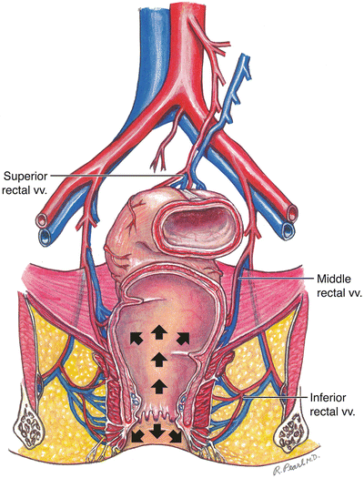
The mucosa proximal to the dentate line lacks somatic innervation. In contrast, the anoderm is richly endowed with cutaneous sensory nerve endings. The blood supply to the rectum and anal canal originates at three levels (Fig. 2.3). The superior rectal artery, the principal blood supply to the upper and middle portion of the rectum, begins as the terminal branch of the inferior mesenteric artery, where it crosses over the left common iliac vessels. As it descends within the sigmoid mesocolon, it bifurcates at the level of the third sacral vertebra into right and left branches that course along and within either side of the rectal wall. Each vessel divides further so that an average of five small mucosal arteries terminate at the level of the anal valves. There appears to be a paucity of anastomoses on the anterior and posterior surfaces of the rectum between the two collateral branches of the superior rectal artery.


Fig. 2.3
The blood supply and venous drainage of the rectum and anal canal. The arrows indicate the direction of lymphatic drainage
The middle rectal arteries usually arise from the internal iliac arteries, travel along the anterolateral surface of the lower rectum, traversing Denonvilliers’ fascia, and enter the rectal wall at the level of the anorectal ring. Although the middle rectal arteries do not run within the lateral stalks, accessory branches may occasionally be found coursing through these ligaments.
The inferior rectal arteries enter the posterolateral aspects of the isciorectal fossae as offshoots of the internal pudendal arteries, which run in Alcock’s canals. Each of these arteries divides further into two to four vessels that supply the external and internal sphincter, as well as the lining of the anal canal.
Stay updated, free articles. Join our Telegram channel

Full access? Get Clinical Tree


