Fig. 7.1
Sympathetic and parasympathetic nervous fibers of the pelvis (Copyright © 2007; Jens-Uwe Stolzenburg and Moonsoft)
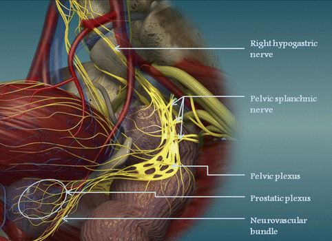
Fig. 7.2
Nerves of the pelvis (Copyright: Moonsoft and Stolzenburg (2007))
The Neurovascular Bundles
The cavernosal nerves, which contain branches of the autonomic nervous system, are responsible for the erectile function. The location of the large percentage of these autonomic nerve fibers is postero-laterally to the prostate, postero-laterally to the sphincter of the urethra and antero-laterally also to the rectum. These fibers can only be recognized by the accompanying vascular structures and are called neurovascular bundles (NVBs). The course of the NVBs is depicted in Fig. 7.3.
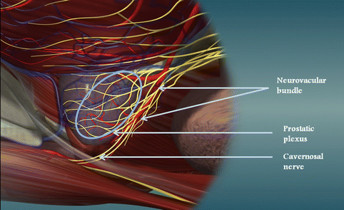

Fig. 7.3
The course of nerves and vessels around the prostate (Copyright © 2007; Jens-Uwe Stolzenburg and Moonsoft)
It is really interesting that the first surgeons who described erectile dysfunction after pelvic surgery were general surgeons that performed surgery to the rectum [17]. Surgeons performing resections of the rectum are following the Denonvilliers’ fascia for the ventral dissection in order to avoid damaging the urinary and erectile function [18]. Walsh and Donker were the first to describe the NVBs and described the surgical technique to preserve these anatomical structures [19]. Each NVB is considered as the structure that contains the autonomic fibers to innervate the corpora cavernosa. It is situated between the lateral pelvic fascia and prostatic fascia (Fig. 7.4). It extends postero-laterally to the prostatic gland. There have been some recent studies showing some variations. A fan-like distribution is found in several cases, rather than a distinct location of the bundle [20, 21]. The further understanding of the pelvic fascias and the location of the NVBs has motivated surgeons to develop new techniques of nerve preservation [22–25] (Fig. 7.5). During the interfascial prostatectomy, the neurovascular bundle is preserved by incising the endopelvic fascia and the periprostatic fascia is excised along with the prostate. In the intrafascial technique, the dissection is performed right on the capsule of the prostate and the periprostatic fascia is not excised with the surgical specimen.
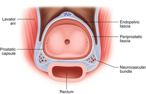
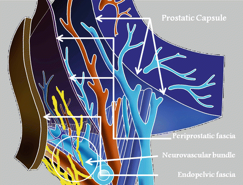

Fig. 7.4
The anatomical landmarks for the preservation of the neurovascular bundles

Fig. 7.5
The prostatic fascias and the neurovascular bundle. The dissection for interfascial nerve preservation takes place between the pelvic fascia and the periprostatic fascia. Thus, the periprostatic fascia is excised with the specimen. The intrafascial dissection takes place between the periprostatic fascia and the capsule of the prostate. The prostate with the capsule is only removed. The neurovascular bundles remain intact in both dissections
An accurate description of the NVB was investigated recently, dividing the NVB in three sections: the anterior section which supplies the cavernosal nerves, the lateral section which supplies the levator ani and the posterolateral section that supplies the rectum [26, 27]. This anatomic segmentation can lead to different surgical approaches in the future, such us partial preservation of the bundle or grafting [16].
Although, the NVBs are considered to carry the nerve fibers for erectile function and their excision results in erectile dysfunction, only a small portion of the patients who undergo non-nerve sparing radical prostatectomy have erectile function postoperatively. A possible explanation is that there is some additional autonomic nerve fibers that innervate the cavernosal body or that there are anastomotic bridges between the pudendal nerves and the cavernosal nerves [28, 29].
The Cavernosal and Pudendal Nerves
The course of the cavernosal nerves is depicted in Fig. 7.6. They are located at a few millimeters from the prostatic capsule at the 5 and 7 o’clock positions. The medial branches supply nerve fibers to the external urethral sphincter, while the lateral branches supply the corpora cavernosa. The course of the pudendal nerves is seen in Fig. 7.7. It is a somatosensory nerve that innervates the bulbospongiosus muscle, the ischiocavernosus muscle and the striated component of the external urethral sphincter. This nerve is often injured during vaginal delivery or pelvic bone fractures [16].
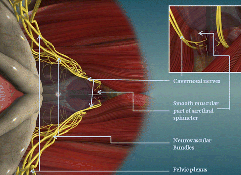
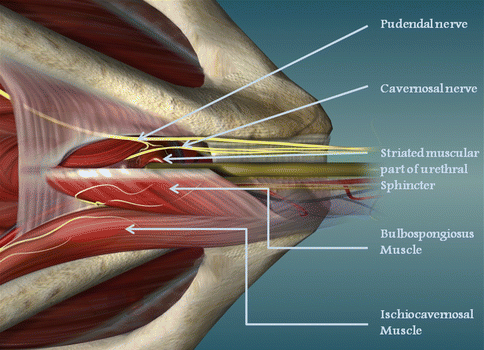

Fig. 7.6
The course of the nerve fibers towards the corpora cavernosa (Copyright © 2007; Jens-Uwe Stolzenburg and Moonsoft)

Fig. 7.7
The pudendal nerve and its motor branches to pelvic muscles (Copyright © 2007; Jens-Uwe Stolzenburg and Moonsoft)
The External Urinary Sphincter
The autonomic innervation of the urethral sphincter is not well defined. Nerve fibers are supplied by the NVBs, while other branches derive from the pudendal nerves [30]. It seems that the urethral sphincter in males, unlike to females, is independent of the muscle tone of the pelvic floor [31]. The distance of the point where the pudendal nerves enter the muscle differs from 3 to 13 mm [32]. The external sphincter has two parts: the external striated segment and the internal smooth muscle segment. For the first segment, the term “rhabdosphincter” is widely used. The posterior part of the striated muscle is interrupted by a tendinous median dorsal raphe [33]. The reconstruction of this dorsal raphe of the posterior rhabdosphincter may increase the early recovery of continence after radical prostatectomy [34, 35]. The internal segment of the sphincter can be divided also to a layer with the muscle fibers oriented in a circumferential plane and another in the longitudinal plane [31]. Continence should probably be attributed to the contraction of external sphincter with the smooth component to provide “basic” continence and the striated component to provide continence during “stress conditions” such as coughing. In addition, the sphincteric system is supported by the surrounding musculofascial structures of the pelvic diaphragm [12, 36, 27].
7.9 Conclusion
The development of radical pelvic surgery led to the further investigation of the pelvic anatomy, in an attempt to prevent the several complications of pelvic procedures. Special interest has been shown to the neuroanatomy of the pelvis, as the autonomic nervous system plays a significant role in the bladder, rectum and erectile function as well as urinary continence. A major breakthrough was the description of the NVBs [9]. The nerve-sparing technique significantly improved the recovery of erectile function. Several refinements of surgical technique have been presented in the recent years in an attempt to achieve earlier recovery of urinary continence. All these techniques are based on the recent knowledge on the anatomy of the pelvis. Further investigations are needed for the complete depiction of the structural and functional anatomy of the pelvis and improvement the surgical techniques.
Key Points
The anatomy of the pelvis is important not only to urologists but also to gynecologists and general surgeons. A multidisciplinary concept of pelvic surgery is of fundamental importance.
The five tissue lines of the umbilical ligaments are the anatomical landmarks for orientation during laparoscopic procedures in the pelvis.
The inguinal rings, external iliac and epigastric vessels are also additional landmarks. The inferior epigastric vessels demarcate direct from indirect inguinal hernia.
The male pelvis is narrower than the female pelvis.
The pelvic plexus is prone to injury in radical cystectomy, antireflux ureteral surgery and rectal surgery.
The nerve fibers of the cavernosal nerves are mainly supplied by the neurovascular bundles which are located posterolaterally to the prostate.
The interfascial technique of nerve-sparing includes the periprostatic fascia in the excision, while the intrafascial technique preserves the periprostatic fascia.
An anatomical segmentation has been presented recently. This may lead to partial preservation of the neurovascular bundles in the future.
Stay updated, free articles. Join our Telegram channel

Full access? Get Clinical Tree


