Type of incontinence
Frequency
Never
Rarely
Sometimes
Usually
Always
Always
Solid
0
1
2
3
4
4
Liquid
0
1
2
3
4
4
Gas
0
1
2
3
4
4
Wears pad
0
1
2
3
4
4
Lifestyle changes
0
1
2
3
4
4
9.4.2 Examination
Detailed anorectal examination is mandatory and may reveal soiling of garments, scars of previous surgery, size of a gaping patulous anus and sphincter tone at rest and sphincter squeeze pressure, reflex contraction when performing digital examination, and fibrosis of the anal canal and adjacent tissue. Also one should look for the presence of a rectocele or a loose rectovaginal septum, perineal descent in response to straining or coughing with opening of the anal canal, and complete rectal prolapse.
9.4.3 Investigations
Clinical evaluation as suggested by Hughes should be adequate enough to assess the cause and the severity of the condition allowing the appropriate plan of action (Hughes et al. 1984).
9.4.3.1 Manometry
Manometry studies will confirm the clinical findings. They are useful in evaluating disorders selectively affecting smooth or striated muscles. These studies are useful particularly if biofeedback training is planned. Furthermore, they allow precise evaluation of postoperative results. Anal manometry determines the sphincter pressures, sensation, rectal compliance, and anorectal reflexes. It is measured by placing a balloon into the rectum which detects the pressure changes. Manometers use air, water perfusion systems, or chips to sense the pressures.
Many different techniques are available, including water-filled perfusion catheters, water-filled or air-filled balloons, sleeve catheter, and pressure transducers. Microtransducers are the most reliable catheters because they minimize the distention of the anus; however the cost and the fragility of these devices restrict their use. The most commonly used sensory devices are 4 to 8 mm in diameter, water-perfused, soft, plastic, and multichannel catheters with radial array.
Squeeze pressure can be measured by asking the patient to contract the sphincter as the catheter is positioned in the pressure zone. Normal values of both resting and squeeze pressures vary among patient population.
Rectal sensory testing includes volumetric measurement of first detectable sensation, sensation of fullness, and the maximum tolerated volume by balloon distension. Hypersensitivity can be seen with inflammatory conditions and poor rectal compliance.
Compliance is measured by inflating the rectal balloon with increasing volume of air or water. The result is expressed as the ratio of the pressure to the volume (C = P/V). The compliance decreases with inflammation, fibrosis, drugs, or surgery.
The normal resting anal pressure is 40 mmHg, and squeeze pressure is 80 mmHg, and these are predominantly the functions of the internal and external anal sphincters, respectively. The compliance refers to the change in volume of the rectum in relation to the change in p. Compliance can change after surgical resection of the rectum or irradiation.
The saline perfusion system has 16 channels for sensing the pressure changes. The balloon is inflated manually to the desired volume (Figs. 9.1 and 9.2).

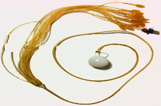
Fig. 9.1
Anal manometry setup: high-resolution impedance manometry system. Electronic chips are used for signal capture (Diagram courtesy of Dr. Uday C. Ghoshal)
Fig. 9.2
Saline perfusion system (Diagram courtesy of Dr. Uday C. Ghoshal)
The lower part of the graph produced by the saline perfusion system corresponds to the anal canal, and the upper pressure corresponds to the rectum (Fig. 9.3).

Fig. 9.3
Saline perfusion system showing the anorectal pressures. There are 16 sensors at a distance of 1 cm, each which tracks the pressure changes in the anorectum (Diagram courtesy of Dr. Uday C. Ghoshal)
9.4.3.2 Measurement of Sphincter Strength
A method for quantitative evaluation of sphincter strength has been described by Henriksen and Huthouisen (1972). A 2 cm diameter ball is inserted into the rectum. The force which is necessary to withdraw the ball is measured.
9.4.3.3 Anal Sphincter Electromyography (EMG)
It checks the health of the pelvic floor, muscles, and the nerves that control the muscles. The average amount of electrical activity when the person relaxes quietly, squeezes to prevent a bowel movement, and strains to have a bowel movement shows whether there is damage to the nerve that controls the external sphincter and pelvic floor muscles are recorded by EMG (Fig. 9.4). It measures the electrical activity of the striated muscles, which includes the external anal sphincter and puborectalis. There are different methods of performing EMG. It can be simultaneously used with videoproctography to detect the electrical activity of the muscles.

Fig. 9.4
EMG study
The role of EMG in the treatment of anal incontinence has reduced with the introduction of EUS. The results of EMG do not really predict the outcome of the sphincter repair. Today, EMG seems to be relevant for sphincter mapping prior to the repair of anorectal malformations and for biofeedback therapy for nonrelaxing puborectalis syndrome.
EMG is used to record electrical activity in the muscle of continence during anorectal function. It is used to determine whether there is evidence of inappropriate puborectalis contraction during defecation. It is also used for sphincter mapping, especially in patients with ectopic anus and congenital anomalies and after severe disruption. Needle EMG either with concentric needle or with single-fiber electrodes had in the past an important role in the clinical mapping of the EAS. The result of needle EMG agrees well with endoanal USG and with surgical or histological methods to identify the sphincter damage. Surface EMG with an anal plug within the anal canal is without pain and with less risk of infection and has a definite role in indicating and applying biofeedback training.
9.4.3.4 Anal Ultrasound
Endoscopic ultrasound (EUS) is one of the latest additions to the armamentarium for the evaluation of the anal sphincter. EUS maps the defects of the internal and external sphincters. It is the single most important investigation in the management of sphincter injuries. The internal and external anal sphincters are seen as hypoechoic and hyperechoic structures, respectively, in the EUS. The puborectalis muscle is seen as a hyperechoic U-shaped structure. The defect in the sphincter could be demonstrated preoperatively, which helps in planning the incision and repair. EUS cannot be performed if there is anal stenosis. The lack of widespread availability and training is also a limiting factor (Fig. 9.5).

Fig. 9.5
Anorectal USG endoscopic ultrasonogram showing the defect in external anal sphincter (Diagram courtesy of Dr. Preveer Rai)
When performed by experienced clinician, endoanal USG approaches 100 % sensitivity and specificity in identifying the sphincter defects. A 15 mm diameter probe with 360° rotation 10 MHz transducer are used to image the sphincter at several levels in the anal canal. The IAS is imaged as a hypoechoic ring close to the transducer, surrounded by a hyperechoic ring representing the EAS. Sphincter defect is measured as a lateral breaks in the sphincter. Endo USG sphincter abnormalities are seen in 90 % of the women whose sole risk factor for fecal incontinence is obstetric trauma.
9.4.3.5 Balloon Proctography and Defecography
This study is useful in establishing any alteration in the anatomical structures and mechanisms of defecation (Macleod 1979; Mahieu et al. 1984; Pichrell et al 1959). It shows how well the person can hold and evacuate stool. It also identifies structural changes in the rectum and anus such as rectoceles and rectal prolapse.
Defecography or the dynamic proctogram is used to define the anatomy and changes of the pelvic floor muscle position with defecation. It can identify abnormalities such as prolapse, perineal descent, and intussusception. This procedure utilizes the paste of barium and potato powder to stimulate the fecal material. Video recording of straining, squeezing, and defecation into a radiolucent commode allows real-time assessment of anatomic changes during defecation.
9.4.3.6 Magnetic Resonance Imaging (MRI)
It is an alternative to anal ultrasound and provides more detailed information about the anatomy especially about the external sphincter (Fig. 9.6).

Fig. 9.6
MRI pictures of the pelvic floor and anorectum
9.4.3.7 Endoscopy
Flexible or rigid sigmoidoscopy and flexible colonoscopy may be useful to exclude colonic pathology.
9.4.3.8 Pudendal Nerve Motor Latency (PNML)
The pudendal nerve, containing fibers from sacral nerves S2–S4, provides motor innervation to the external sphincter and receives sensory information from the perineum. PNML measures the conduction time to external sphincter contraction after the nerve stimulation at the level of the ischial spine. This is easily performed using a digitally mounted device with a stimulating electrode mounted at the fingertip and recording electrode mounted at the base of the finger. The mass production of a self-adhesive disposable electrode, which can easily be mounted on a gloved finger, has enabled this assessment to become routine in most centers.
The normal latency is 2.1 ± 0.2 ms. Prolonged latency may be associated with obstetric injury, perineal descent, prolapse, and medical neuropathies The success rate after sphincter repair decreases from about 90 % with intact pudendal nerve to 50 % in the presence of pudendal neuropathy.
9.5 Treatment of Anal Incontinence
9.5.1 Conservative Treatment
9.5.1.1 Diet
Dietary modification is important for successful management. Both diarrhea and constipation can contribute to incontinence. So dietary advice must be tailored to address the underlying cause, or it may be ineffective or counterproductive. In persons with disease aggravated by diarrhea or those with rectal loading by soft stools, the patients must be advised to increase dietary fiber and reduce intake of wholegrain cereals/bread, fruit, and vegetables which contain natural laxative compounds (rhubarb, figs, prunes/plums); beans, pulses, cabbage, sprouts, and spices (especially chili); artificial sweeteners (e.g., sugar-free chewing gum); alcohol (especially stout, beer, and ale); lactose if there is some degree of lactase deficiency; and caffeine. Caffeine lowers the resting tone of the anal canal and also causes diarrhea. Excessive doses of vitamin C, magnesium, phosphorus, and/or calcium supplements may increase fecal incontinence. Reducing olestra fat substitute, which can cause diarrhea, may also help (Norton et al. 2007).
9.5.1.2 Pharmacological Treatment
Patients with minor degree of incontinence or patients who are unfit for surgery may be treated conservatively (Brocklehurst 1978; Marti and Noethiger 1981). Stool thickeners, bulk-forming agents, and high fiber intake should be routinely prescribed to obtain firm stools. Evacuation can be stimulated by glycerin suppositories used at predictable time of the day. The suppositories produce rectal distention. Through repeated applications, rectal volume may increase and the sensations of rectal distention are stimulated. The patient will then be continent until the next artificially induced bowel movement. Various pharmacological agents are:
(a)
Adsorbents:
Kaopectate – useful for mild degree of incontinence and acts by absorbing excess fluid in the stool.
Opium derivative – loperamide (Imodium), commonly used. Others agents are diphenoxylate hydrochloride (Lomotil), diphenoxylate hydrochloride + atropine, codeine, and tincture of opium.
(b)
Tricyclic antidepressant:
Like amitriptyline (20 mg, daily). It has anticholinergic and serotoninergic properties.
(c)
Bulking agents:
Better used for patients of diarrheal variety of irritable bowel syndrome
(d)
Bile salt binders:
Cholestyramine and colestipol. These resins treat bile acid diarrhea by binding with bile salts in the small intestine.
(e)
Topical agents:
Act on the internal sphincter and increases the resting tone, e.g., 10 % phenylephrine
9.5.1.3 Bowel Management
The aim is to allow the patients to produce a complete bowel movement at a schedule time by using an individualized combination of dietary measures, laxatives, suppositories, enemas, and digitization. This method is useful for patients with neurological problems, diabetes, and congenital anorectal malformation. This is also useful for patients with overflow incontinence and pediatric patients with encopresis having symptoms of seepage of stool from full rectum. The aim is complete cleansing of the colon by the use of various combinations. Later, the use of the daily formulation of polyethylene glycol/laxative is better.
9.5.1.4 Physical Treatment
Muscular training is very important to stimulate muscles and to increase the muscular activity by regular sphincter exercise. Training may be voluntary, with or without biofeedback control of the increased endoanal pressure level, or may be performed by electrical stimulation using implanted electrodes or externally activated plugs (Bleijenberg and Kuijpers 1987; Loygue and Dubois 1964).
Electrical stimulation seems not to result in an increase of anal tone but regression of muscular fatigability. If contractions can be sustained for 50–60 s, rectal compliance will increase and then continence can be achieved.
9.5.1.5 Biofeedback
Biofeedback has a broad acceptance as a useful treatment of fecal incontinence. The term “biofeedback” describes a therapeutic instrument that derives from psychologic “theory of learning.” This type of learning is also called instrumental learning or operant conditioning. A body function that cannot or can only be perceived poorly by the subject under normal condition is measured by a technical device and demonstrated (feedback) to the subject. There are two methods of biofeedback training:
(1)
Response to rectal distension using manometric techniques
(2)
Unrelated to rectal distension, muscle strengthening using EMG or manometric technique
Biofeedback may result in a better coordination of sphincter activity in carefully selected (without any obvious cause on defecography but abnormal manometry) and motivated patients (Corman 1985; Denis et al 1983). A balloon placed within the anal canal is connected to a transducer, and a graph on the monitor will show the pressure readings at different stages of attempted defecation. The patient observes the anal pressure peak reached by sphincter contraction with or without rectal distention. He is trained accordingly as when and how to relax the sphincters. Improvement usually occurs within three to five training sessions.
The treatment is supervised by a biofeedback therapist who evaluates patients during 6–8 weeks. Both methods are effective, although some patients may respond better to one system than the other. Biofeedback therapy is best applied to motivated patients with some ability to voluntarily contract the external anal sphincter (even if the muscle is partially disrupted) and an intact rectal sensation. Biofeedback is beneficial in patients with incontinence of variable etiologies such as diabetes, after childbirth, and after anorectal surgery; however, the best results can be seen in patients who have primarily a sensory problem in the anal canal leading to insensible loss of feces. The results of biofeedback training in different patient groups suffering from incontinence vary from 64 to 89 %, with an overall success rate of approximately 70 %. Symptom improvement may sustain several years after treatment. The exact mechanism of success is unclear. Nevertheless, it is safe and effective and does not preclude other treatments. Biofeedback may result in a better coordination of sphincter activity in carefully selected (without any obvious cause on defecography but abnormal manometry) and motivated patients (Corman 1985; Denis et al 1983).
9.5.1.6 Faradic Stimulation
The goals of this treatment are to increase the strength of the pelvic floor muscles and normalize the reflex activity of the muscles via stimulation of the afferent and efferent fibers of the anorectal sphincters. An electric muscle stimulator with sinusoidal, faradic, and interrupted direct current outputs is used. It has been suggested that electrical stimulation can transform type II (fast twitch) motor units to mainly type I (slow twitch) units (Sokunbi and Okunsanya 2002). Type I motor units have been observed to generate more tension, thereby increasing the tone of the pelvic floor muscles, resulting in decrease in the symptoms of incontinence. Modifying sensations around the anorectal region can inhibit fecal incontinence by stimulating the afferent fibers, whereas stimulation of efferent fibers can induce voiding by stimulating the contraction of the region.
The patient lies in a prone position with pillows under the abdomen and the ankle. The patient should be adequately draped to allow minimal exposure of the area being treated. A four-pole electrode (6 cm by 8 cm) application is utilized with two electrodes placed medial to the ischial tuberosities on either side of the anus, and the remaining two are placed along an imaginary line joining the posterior inferior iliac spine, 5 cm each from the midline (Continence Foundation 2001; Sokunbi and Okunsanya 2002).
The duration of the stimulation is between 20 min and an hour per day for about 20 days. The treatment may be needed for a period ranging from a few weeks to several months (Continence Foundation 2001; Sokunbi and Okunsanya 2002).
9.5.1.7 Percutaneous Tibial Nerve Stimulation (PTNS)
There is extensive evidence regarding the efficacy of PTNS in urinary incontinence. Data on the efficacy of PTNS for fecal incontinence (FI) are limited to a small case series with short follow-up. However, in a recently cited study, it was found to improve the FI score after 12 sessions of treatment and further after top-up sessions.
PTNS was well tolerated with high acceptability in the majority of patients. The effect of PTNS diminishes with time (usually after 42 months), and additional therapy sessions at 6 months’ intervals may result in greater improvement (Hotouras et al. 2014).
9.5.2 Surgical Treatment
The main indication for surgical intervention is major incontinence refractory to biofeedback and sacral nerve stimulation. This is the first line of treatment option in cases of obstetric injury with isolated sphincter defect and has an excellent outcome.
The choice of surgical techniques depends mainly on the nature and the level of the lesion responsible for incontinence. Bowel preparation and sterilization are mandatory.
The aims of the various surgical techniques used are reduction of anal canal diameter, sphincter construction, reinforcement of the occlusion mechanism, increase of muscular mass, decrease in the size of the anorectal angle at rest, substitutive sphincteroplasty, and artificial sphincter implantation.
9.5.2.1 Thiersch Operation
Encirclement of the anal orifice with wire or nonabsorbable suture material, fascia lata, or Teflon has been used to prevent rectal prolapse (Goebell 1927). This procedure creates a static barrier to passage of rectal content, especially solid feces, but not liquids or flatus. It does not contribute anything to voluntary control and maintenance of continence. This procedure is frequently complicated by secondary infection and extrusion of the suture material as a foreign body and is offered to old and debilitated patients and children (Fig. 9.7).

Fig. 9.7
Thiersch operation
9.5.2.2 Repair of Obstetrical Injuries
In this type of injury, the posterior vaginal wall, muscles of the perineum, the external anal sphincter, the internal anal sphincter, and the wall of the rectum are torn. Hence it is necessary to stitch back all these components to regain the full continence. An attempt at repair should be made at the time of delivery. If this fails, at least 6 months should elapse before an attempt at repair is made which gives the tissue sufficient time to return to normal. The operation at this time will be easier to perform and chances of success are enhanced.
Layer method of repair is done for old obstetric tears (Fig. 9.8a–e). An inverted semilunar incision is made at the junction of the posterior vaginal wall and the rectal mucosa, and the lateral end of the incision should reach the stumps of the sphincters. The vaginal flap is dissected upward keeping close to the vaginal wall. The dissection is carried out till the puborectalis is demonstrated on either side. The dissection is then carried out to demonstrate and mobilize the stump of the sphincter muscle, which is then grasped by Babcock forceps. The deficient seromuscular layer of the rectum is repaired by taking interrupted sutures with atraumatic 2-0 PDS. The puborectalis part of levator is stitched in front by two–three interrupted stitches. The stumps of mobilized sphincter ends are then stitched together in front with same suture material or Prolene. Excessive vaginal mucosa is excised and the wound is closed with a drain, if necessary. An indwelling urethral catheter is passed into the bladder and retained for 4–5 days; the drain is removed after 48 h and skin suture after 7 or 8 days. It is not necessary to constipate the patient in immediate postoperative period. Stool softeners are given from the third day of operation to have an easy passage of soft stool. The addition of an antibiotic is essential for the prevention of infection.


