Fig. 11.1
Guidewires (Straight tip and Angle Tip, both Boston Scientific Glidewire)
More recently, hybrid guidewires have been introduced, which combine the beneficial properties of multiple types of wires (e.g. Sensor Wire™, Boston Scientific; UroWIRE XF™, Applied Medical; and U-Nite™, Bard Urological). In general, these wires possess a kink-resistant core, a distal tip with hydrophilic coating, and a soft proximal tip for backloading of a ureteroscope [2].
Comparisons of Wires
Given the variety available on the market, several groups have attempted to compare wire characteristics in order to help guide selection of the appropriate wire. Clayman et al. provided an early systematic comparison of guidewires, assessing two main functions – safe access to the upper tract and providing a guide over which to advance instruments [6]. The Boston Scientific Glidewire™ was suggested as the safest access wire, as it demonstrated the most lubricity and the safest tip. With respect to the ability to guide other instruments, the Amplatz super stiff guide wire demonstrated the greatest shaft rigidity. The rigidity of this wire was significantly greater (p < 0.05) than all other wires tested.
In a comparison of hydrophilic wires only, Torricelli et al. compared wires with hydrophilic coatings on a variety of similar parameters [8]. Similar to the Clayman study, the Boston Scientific Glidewire™ was declared the safest wire to avoid perforations, secondary to the most flexible tip and highest force required to perforate a test sheet of aluminum foil. Notably, the Boston Scientific Zipwire™ had similar results with respect to tip perforation (p = .340 versus the Glidewire), but the higher shaft rigidity of the Glidewire (p < 0.001 versus the other tested wires) suggested improved versatility for use with other instruments. It has been suggested that the very low coefficient of friction with hydrophilic coating may decrease tactile feedback and enable creation or propagation of a false passage.
Sarkissian et al. also provided a systematic evaluation of the hybrid guidewires [9]. Noteworthy is their comparison of shaft stiffness to the Amplatz SuperStiff and tip hydrophilicity relative to the NiCore™ and RadiFocus™ guidewire. They found that the Amplatz wire remained the stiffest wire available, with at least 34 % greater shaft bending force than the other wires, and accordingly, it continues to be the wire most suited to passing instruments. All in all, the wires produced by Boston Scientific (RadiFocus™, Sensor™, Amplatz) were noted to have lower tip bending force compared to the Bard-manufactured wires (U-Nite™, Ni-Core™), which confers greater applicability in the difficult ureter. Further, the RadiFocus™ wire showed the highest force required to puncture tin foil (p < 0.001), and thus may be considered as one of the safest hybrid wires for initial access. Finally, it was noted that the Sensor™ wire had the highest coefficient of friction (p < 0.0002), supporting its role as a safety wire with low risk of inadvertent migration.
While the above studies were performed in laboratory simulations, Liguori et al. tested several guidewires within cadaveric ureters [10]. As with other studies, the nitinol hydrophilic wires (Terumo Radifocus™, Boston Scientific Sensor™) were considered the safest for initial access. In fact, these two wires never perforated the cadaver ureter. Further, the Sensor™ had the lowest friction value of the tested wires, and was concluded to be the safest. Comparison was also made of several PTFE-coated wires, which would be considered more appropriate for passage of catheters or stents. Among those considered (Boston Scientific PTFE guidewire, Medtronic PTFE guidewire, and Emerald Cordis Guidewire), tip safety and friction tests were similar, but the superior resistance to bending of the Boston Scientific PTFE guidewire suggested it as the most preferable for use with introducing instrumentation.
Ureteral Dilation
After passage of guide wire(s), the ability to introduce further elements such as an ureteroscope, access sheath, or other instruments may be limited by a tight ureteral orifice, ureteral narrowing/stricture, or both. These may be related to normal anatomic variants, congenital abnormality, or related to prior medical treatment or surgery. The most common area requiring dilation is at the ureterovesical junction [2]. Ureteral dilation can be accomplished by coaxial dilators, ureteral access sheaths, balloon dilation, or interval ureteral stenting.
Coaxial dilation was the original method of ureteral dilation. This is accomplished by sequential passage of increasingly large dilators or a single tapered dilator. Both are passed over a guidewire to promote safe dilation. The use of a stiff shaft wire provides the best guide for passage of coaxial dilators. The main advantages of coaxial dilation include relative quickness of dilation as well as significantly lower cost compared to balloon dilation [11]. However, the dilation occurs via axial shearing force, which may cause mucosal injury and predispose the ureter to strictures [2, 11].
Ureteral access sheaths (UAS) can be considered a variant of coaxial dilation. These will be discussed in detail elsewhere in the chapter, but consist of an outer sheath with an inner tapered dilator. Together, these function similarly to a tapered dilator, but the inner portion is able to be removed, leaving the outer sheath for passage of ureteroscopes or other instruments. As the UAS is passed, the ureter is dilated similarly to with other coaxial dilators. This provides secure access proximal to the area of narrowing after dilation, but is again associated with axial shear forces that may damage the ureteral mucosa.
Conversely, balloon dilators provide radial dilation to the area of narrowing; force is exerted perpendicular to the mucosal axis, causing less mucosal damage [12]. The balloon catheters are between 4 and 7Fr in diameter, and balloon length can range between 2 and 10 cm [2, 3]. The system typically consists of a balloon situated around a central channel, which is used to advance the system over a wire. A second channel consists of tubing to the balloon for balloon inflation. The edges of the balloon length are marked with radiopaque markers ensuring knowledge of the outer limits of dilation with fluoroscopy. The important features to consider with any balloon dilators include the burst pressure as well as the compliance. The burst pressure of most ureteral balloons ranges between 8 and 20 atmospheres of pressure. The rupture of a balloon can cause significant damage to the urinary tract, so these products often package pressure gauges in the kit to ensure safe operation. The compliance of the balloon dilator determines its effectiveness, as a more compliant balloon may not overcome the deformity at the area of the narrowing, creating a classic hourglass appearance on fluoroscopy. The ideal balloon is therefore non-complaint, i.e., it will expand to 100 % of its designed diameter despite external forces. A comparison of ureteral balloons by Hendlin et al. assessed the balloon diameter and pressure with varying external compressive forces. This study demonstrated the Cook Ascend AQ™ balloon to be the most reliable balloon for use, as it reproducibly reached published diameter at the designated burst pressure. Notably, many balloons tested were not able to reach to target diameter even at burst pressure when subjected to larger external forces, which may limit the efficacy of these products [13].
Finally, the concept of interval ureteral stenting deserves mention. While not technically a dilation strategy, placing a ureteral stent during the acute stone episode to return at a later date for URS allows passive dilation. This is associated with significantly less chance of subsequent ureteral dilation. A study assessing predictive factors for balloon dilation during semi-rigid URS demonstrated fewer requirements for dilation in patients with prior stent placement. Overall, dilation was performed in 27.5 % of procedures, while patients with prior stent required dilation only 1.8 % of the time [12]. In the setting of difficult ureteral access, this option may prove to be a safer strategy to avoid iatrogenic injury.
Manipulation Devices
Once the calculus is visualized, the ability to manipulate and/or remove it is dependent on the use of a manipulation device such as basket or forceps. Over time, the size of these devices has decreased enabling their passage through the working ports of most ureteroscopes. Baskets generally consist of several wires configured to surround the calculus to allow for manipulation, while forceps consist of two or more prongs that grasp the stone for manipulation. In general, both baskets and forceps rest within the shaft of the device, and are then extended beyond the shaft toward the target by the user using a handle at the end [14].
There are several characteristics of basket construction that must be considered for selection. As with guidewires, baskets may be composed of stainless steel or nitinol alloy [3]. The stainless steel baskets are stronger and may be preferred in a situation where ureteral distention is necessary for stone grasping. On the other hand, the elasticity of nitinol baskets may prove helpful in snaring eccentrically located stones. Further, the smaller size (in general) of nitinol devices allows for improved visualization through a similarly sized scope.
Another important characteristic is the wire configuration of the basket: flat-wire, helical, or multi-wire (Fig. 11.2). Baskets generally consist of 3–16 wires arranged in various configurations. Flat-wire baskets consist of wires arranged concentrically. This is the most commonly used shape of basket. While it will reliably grab a target calculus, the shape may be less suited for manipulation of multiple calculi [3]. Helical baskets, as the name suggests, consist of wires that intertwine in a helical pattern around the target. This pattern grips the calculus securely, and as such may prove helpful for the entrapped stone, but the smaller openings may limit the size of calculus. Finally, multi-wire baskets consist of several wires arranged parallel to form a “canopy” [3]. This style may be most useful for removal of multiple calculi (Fig 11.3).
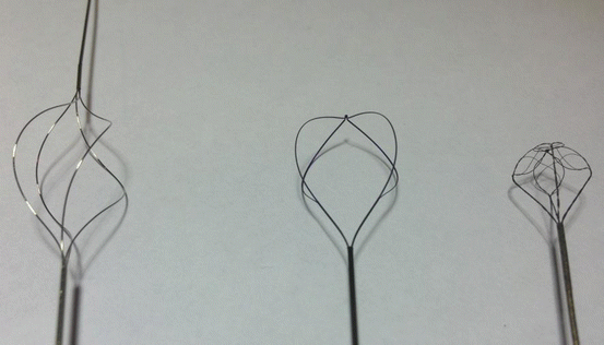
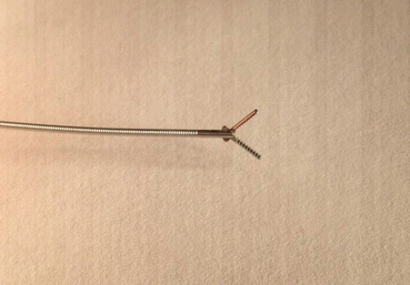
Fig. 11.2
Baskets examples: helical with tip (bard urological), flat (Boston Scientific), Multi-Wire (Cook)
Fig. 11.3
Forceps example
Some surgeons may choose use of a basket with a hollow core. This allows for passage of the basket over a wire or for advancing a laser fiber through the basket. The ability to pass a laser fiber may prove useful for performing lithotripsy on a stone held within the basket. Care must be taken to prevent striking the basket wires and potentially releasing a foreign body in the ureter.
Finally, the presence or absence of a tip is an important decision in basket selection. The tips extend beyond the cage of the basket, and in the ureter may help to negotiate a difficult ureter. However, a tip may prove to be dangerous, as it increases the risk for mucosal damage and/or perforation. As such, most baskets available today are of tipless design. If straightening a tortuous ureter is necessary with the use of tipless baskets, one can advance a guidewire first to assist with this.
Forceps consist of two or more prongs that extend from the shaft to grasp the target. They are different from baskets in that the prongs do not converge to a single point distally. Forceps may potentially be safer in the setting of larger calculi; i.e., if the stone becomes trapped in the ureter during removal, it may be more readily released, and reduce the risk of ureteral trauma or avulsion [3]. A comparison of various forceps and baskets in a porcine ureter model demonstrated that the simple two-prong forceps provided the fastest extraction for a single ureteral stone. The three-prong graspers were only slightly slower, but produced significantly higher tissue damage on macroscopic evaluation of the tissue. Within the baskets studied, the helical type basket provided the most efficient extraction in all models (single stone, impacted stone, steinstrasse) [15]. Comparison studies are available in the literature which evaluate individual basket types on applicable characteristics [16, 17]. While many baskets were tested, these studies found that the Sacred Heart Halo™ basket exhibited the best radial dilation, while the Cook N-Circle™ was the most expedient to extract a stone.
Ureteral Access Sheaths
The use of flexible ureteroscopes for calculus disease has drastically increased the abilities of the endourologist to treat ureteral calculi. However, difficulties still exist in their use. For example, it is common that complete removal of a ureteral stone may require several “passes” of the scope with a basket or forceps. This increases the chances of urethral or ureteral damage, and decreases the life of the ureteroscope as it is repeatedly passed over a wire. Further, the small irrigation channels of the flexible ureteroscope require significant pressure to allow adequate visualization. This pressure can transmit to the renal pelvis and cause increased pain or fluid absorption secondary to pyelovenous backflow. The ureteral access sheath (UAS) was introduced to address such concerns, and provide consistent, safe access to the ureter and kidney.
While various models exist, the basic design of the UAS remains the same (Fig. 11.4). A tapered dilator rests inside the outer sheath, connecting to provide a smooth, dilating surface. The outer sheath is usually coated with a hydrophilic coating to reduce the coefficient of friction and allow for smooth passage of the UAS. Options with the UAS include outer diameter (ranging from 9.5 to 16Fr) and sheath length (20–55 cm) [18]. The appropriate size selection depends on the caliber of the ureter and ureteral orifice, as well as the size of the ureter and location of the stone to be treated. A UAS is placed over the working wire and advanced under fluoroscopic guidance to the desired location. It is recommended to have a safety wire in place, and introduce the UAS over a stiff working wire to reduce the risk of wire buckling and iatrogenic injury. Once in appropriate position, the working wire and inner obturator are removed, and URS may commence.
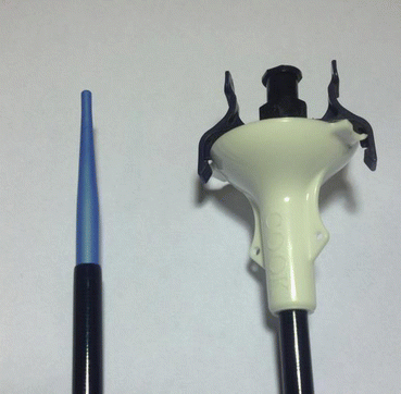
Fig. 11.4
Ureteral access sheath (cook)
Certain advantages and disadvantages exist with the use of the UAS. Certainly, using a UAS in situations with large or multiple calculi will decrease operative time by allowing quick removal and re-entry of the ureteroscope. Prospective trials have compared URS for lithotripsy (with basketing) with and without UAS, and these have shown significantly decreased operative time and cost in cases utilizing the UAS. Operative time was shown to be about 10 min less with the UAS in place, and achieved similar stone-free rates despite increased stone burden in the UAS group [19]. Further, the ability to re-introduce the ureteroscope without assistance of a guide wire reduces trauma to the inner sheath of the ureteroscope. As such, there may be decreased ureteroscope repair cost and repair downtime.
In addition, the UAS decreases the intrarenal pressure generated during URS by providing a pathway for irrigant outflow. The use of high-pressure irrigation systems during URS can be associated with very high intrarenal pressures. The specific effects of this short rise in pressures are not known, but theoretical concerns warrant minimizing pressures when possible. Further, it has been suggested that elevated intrarenal pressure increases pyelovenous backflow and subsequent fluid absorption [19]. Although prior studies have not shown significant fluid absorption, the theoretical risks may exist in patients with cardiac and/or renal dysfunction. One study utilized renal pressure measurements via percutaneous nephrostomy tubes during URS for ureteral calculi. It demonstrated decreased pressures in both the renal pelvis and parenchyma with the use of the UAS [20]. Further, the overall increase in flow of irrigation was associated with subjective improvement in visualization.
Related posts:
 Radiation Exposure to the Patient and the Urologist
Shock Wave Lithotripsy for the Treatment of Ureteral Stones
Selecting the Appropriate Treatment Modality for Ureteral Calculi
Complications in the Treatment of Ureteral Stones: Prevention and Management
Radiation Exposure to the Patient and the Urologist
Shock Wave Lithotripsy for the Treatment of Ureteral Stones
Selecting the Appropriate Treatment Modality for Ureteral Calculi
Complications in the Treatment of Ureteral Stones: Prevention and Management
 Semirigid Ureteroscopy for Ureteral Calculi
 Tips and Tricks in the Treatment of Ureteral Stones
Stay updated, free articles. Join our Telegram channel

Full access? Get Clinical Tree


