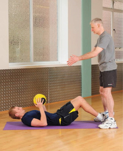
Abdominal Training Following Surgery
Many people tend to think that after surgery on the abdomen they will always have weakness and a ‘floppy tummy’. This is not true. Although a scar is present and the muscles have been affected, it is possible to re-train the abdominal muscles and get full function back in this area.
Surgical operation scars
Following a surgical operation in the abdominal region, you are left with a scar; its size and position will dictate the type and intensity of abdominal training appropriate.
There are many positions for scars and fig. 16.1 shows some of the most common. Essentially an incision is either vertical, travelling down the length of the body, transverse going across at 90 degrees, or oblique, travelling at an angle. The size of the scar will be dependent on the procedure carried out and the structure affected. A lot of surgery is carried out using keyhole procedures; a number of tiny scars are left where the fibre optic camera and small instruments used by the surgeon were passed through.
In most operations a surgeon prefers to cut between (split), rather than through, muscle fibres making healing quicker and stronger. Larger nerves are generally avoided but, because small nerve branches may have been cut during surgery, your scar area may feel slightly numb afterwards.
Vertical incisions
Midline
A midline incision is the most common general approach to the abdomen. It is used because the scar passes through the tissue joining the two large rectus muscles (the linea alba), and so no muscle tissue is actually cut. In the upper abdomen the scar extends from the middle of the lowest ribs (xiphoid process) to just above the tummy button (umbilicus). If more room is needed, the scar may be lengthened by taking it around and then below the umbilicus. With any operation what you see on the surface disguises the amount of tissue that has been cut. To get to an organ, the surgeon must cut through
skin, the fat layer beneath, the muscle (or in this case linea alba) and finally the peritoneum or lining of the abdominal cavity which consists of sheets of connective tissue holding the organs in place.
skin, the fat layer beneath, the muscle (or in this case linea alba) and finally the peritoneum or lining of the abdominal cavity which consists of sheets of connective tissue holding the organs in place.
Keypoint
With a surgical incision, skin, fat, fascia and muscle may be cut and blood vessels sealed before the target organ is even touched.
Paramedian
A paramedian incision is parallel to the midline approach above, but about 3 cm from the midline. More tissue is cut through now, because the surgeon must cut into the covering layer of the rectus muscle, the rectus sheath. The rectus muscle fibres are divided rather than cut. The nerves and blood vessels on the outer (lateral) side of the muscle can be affected by a paramedian incision, so the muscle tends to appear wasted after surgery and take more time to build back up.
Transverse and oblique incisions
Subcostal
The subcostal or kocher incision is often used for gall bladder operations. As it is performed at the top end of the abdomen, less tissue is cut through, making it useful for an obese individual. With this incision, the rectus abdominis, internal oblique and transversus abdominis muscles are all cut and any small blood vessels that bleed will be cauterised (sealed with heat), so the muscles will take more time to recover. Often a small nerve (the eighth thoracic nerve) may be cut as well, delaying muscle recovery slightly.
McBurney
The McBurney incision is most commonly used for appendix operations. It is made about 2 cm above (proximal) to the sharp front edge of the pelvis (the anterior superior iliac spine) on the right side of the body. The incision runs parallel to the fibres of the external oblique muscle fibres, so these muscles are separated and retracted rather than cut, speeding post-surgical recovery.
Lower uterine (bikini)
The lower uterine incision is normally used with a caesarian section (c-section), and is made across the lower abdomen, just above the top of the bladder. Sometimes a midline (vertical) incision may be used if the baby is very large. C-sections are used in about 20 per cent of births in the UK. This procedure is normally carried out using regional anaesthesia – that is an epidural or spinal painkiller where the mother remains awake. Again, muscles are not cut, but the rectus muscles are separated and stretched. Recovery usually only takes about four days.
Keypoint
Where muscle fibres are separated rather than cut through, recovery will be quicker.
Hernia
A hernia occurs when tissue presses through a weakened area of the body, and hernias can occur in many body regions. For example, you can get a hernia on the back of the knee (Baker’s cyst) where the knee joint capsule presses through the gap between the muscles at the back of the knee. However, most people think of the abdominal region when hernia is mentioned, and we will focus on this area. There are several types of hernia, the most common being the inguinal hernia.
Inguinal hernia
An inguinal or ‘groin’ hernia is the most common type of hernia in adults. It is seen more frequently in men and occurs due to weakness in the abdominal wall. The hernia presses through a naturally weak region in the lower abdomen called the myopectineal orifice (MPO). This small area has no strong muscle tissue reinforcing it. Above the MPO is an arch formed from fibres of the internal oblique and transversus muscle. Medially (towards the centre of the body) is the rectus abdominis muscle, with its covering sheath, and laterally (to the outside of the body) lies the iliopsoas muscle. At the bottom of the MPO is the pectineal ligament near the pubic region of the pelvis.


