and George Velmahos1
(1)
Division of Trauma, Emergency Surgery, and Surgical Critical Care, Massachusetts General Hospital, Harvard Medical School, Boston, MA 02114, USA
Abstract
The vast majority of traumatic pulmonary injuries can be treated nonoperatively or with tube thoracostomy alone. The main indications for operative intervention are hemorrhage or large airway injury. Major pulmonary hemorrhage is most commonly caused by penetrating mechanisms, while large airway injury may result from blunt or penetrating mechanisms. Lung injuries may also be found at the time of thoracotomy performed primarily for management of other injuries. This chapter discusses the indications for surgical management of lung injuries, describes techniques for obtaining vascular control and addressing parenchymal and airway injuries, and explains basic considerations in postoperative care.
6.1 Introduction
The vast majority of traumatic pulmonary injuries can be treated nonoperatively or with tube thoracostomy alone. The main indications for operative intervention are hemorrhage or large airway injury. Major pulmonary hemorrhage is most commonly caused by penetrating mechanisms, while large airway injury may result from blunt or penetrating mechanisms. Lung injuries may also be found at the time of thoracotomy performed primarily for management of other injuries. This chapter discusses the indications for surgical management of lung injuries, describes techniques for obtaining vascular control and addressing parenchymal and airway injuries, and explains basic considerations in postoperative care.
6.2 Initial Evaluation and Indications for Surgery
Patients with lung trauma require the same measures for initial stabilization as other trauma patients including airway control and a thorough evaluation for other injuries. This is not discussed at length here. Although there are some considerations for airway injury discussed below, the primary means or airway management remains rapid-sequence orotracheal intubation with a single-lumen endotracheal tube in patients who cannot oxygenate or ventilate adequately on their own. Other chapters on neck and mediastinal trauma discuss special considerations in tracheal or upper airway injury.
All patients with chest injury should be evaluated for hemothorax or pneumothorax. Essentially all patients with operative lung injuries will present with one or both of these findings. This assessment can be made in the trauma bay with chest X-ray or, if the clinician is adequately experienced, with ultrasound as part of the extended focused abdominal sonography for trauma (E-FAST) exam. In unstable patients with signs of chest injury on physical exam, urgent tube thoracostomy can be both diagnostic and therapeutic and should not be delayed to perform imaging. Whether performed before or after imaging, all patients with significant pneumothoraces or hemothoraces after trauma should undergo tube thoracostomy (Fig. 6.1). Even for urgent tube thoracostomy, correct sterile technique with wide draping and gown, glove, and mask use can almost always be adhered to. Additionally, we recommend a single periprocedural dose of antibiotics.
Fig. 6.1
Algorithm for the initial management of lung trauma
The primary indications for thoracotomy in patients with suspected lung injury are major bleeding or major airway disruption. Immediate drainage of over 1,500 mL of blood or greater than 200 mL of blood per hour for 4 h after tube thoracostomy is an indication for thoracotomy in the case of bleeding, as this degree of hemorrhage is unlikely to resolve without surgical intervention. No further preoperative evaluation is needed (Fig. 6.1).
In the case of major airway injury, persistent pneumothorax despite adequate chest tube placement, a blowing air leak, loss of tidal volumes in mechanically ventilated patients, or a “fallen lung” or pneumomediastinum on chest X-ray are signs of major airway injury (Fig. 6.2). Occasionally, placing a chest tube to suction under these conditions will worsen the clinical situation, as it can evacuate the entire tidal volume and interfere with ventilation of the uninjured lung. If this occurs, the tube should be placed to water seal. Flexible bronchoscopy remains the standard for diagnosis of large airway injury – it allows identification of the site of injury and is the most sensitive means of excluding airway injury when the clinical picture is not clear. While standard chest computed tomography (CT) may demonstrate a site of airway injury and is useful to evaluate for associated injuries, it is not sensitive and cannot reliably exclude airway injury.
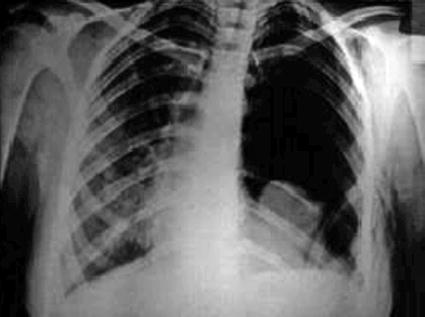
Fig. 6.2
Chest X-ray showing a “fallen lung” suggesting a major bronchial injury
In the absence of these signs of major bleeding or airway disruption, tube thoracostomy alone suffices for treatment of pneumothorax or hemothorax. This is the case in over 90 % of traumatic lung injuries.
6.3 Operative Preparation and Choice of Incision
For patients in whom injury can be confidently isolated to one hemithorax and who are stable enough to allow for positioning, a posterolateral thoracotomy through the fifth intercostal space with the patient in the lateral decubitus position provides the best exposure for surgical treatment of pulmonary injuries as it allows all surfaces of the lung to be exposed and allows anterior and posterior exposure of the pulmonary hilum. However, victims of both blunt and penetrating lung trauma are at high risk for extrathoracic injuries that may require simultaneous management, and these considerations will necessarily influence the approach to the thoracic injury. For example, a patient requiring a simultaneous laparotomy will need to be placed supine, making a lateral or posterolateral thoracotomy impossible. Many pulmonary injuries can be definitively managed or at least damage controlled through an anterolateral thoracotomy. Occasionally these patients may need to be repositioned after extrathoracic injuries have been addressed to allow definitive repair of their thoracic injuries.
A double-lumen endotracheal tube is not necessary for the majority of lung trauma cases, but a contralateral double-lumen tube is especially useful, if possible, for cases of suspected or confirmed main stem or lobar bronchial injury.
6.4 Techniques for Hemorrhage Control
Thoracic exploration for hemorrhage is most often performed on the basis of hemorrhage from a thoracostomy tube. The site of bleeding is seldom localized preoperatively. A thorough exploration must be performed. Techniques for addressing major vascular injuries (such as cardiac, aortic, or great vessel injury) are described elsewhere in this book. Bleeding from chest wall sources – usually either an intercostal artery or the internal mammary artery – can be controlled by ligation.
Related posts:


Stay updated, free articles. Join our Telegram channel

Full access? Get Clinical Tree


