Fig. 20.1
Scanning of a transabdominal ultrasound phantom (Courtesy of Kyoto Kagaku Co., Ltd., Kyoko Japan)
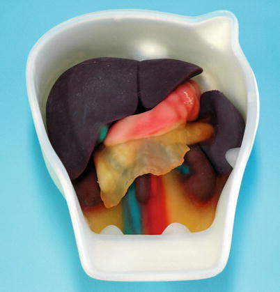
Fig. 20.2
Intraoperative/laparoscopic ultrasound phantom (Courtesy of Kyoto Kagaku Co., Ltd., Kyoko Japan)
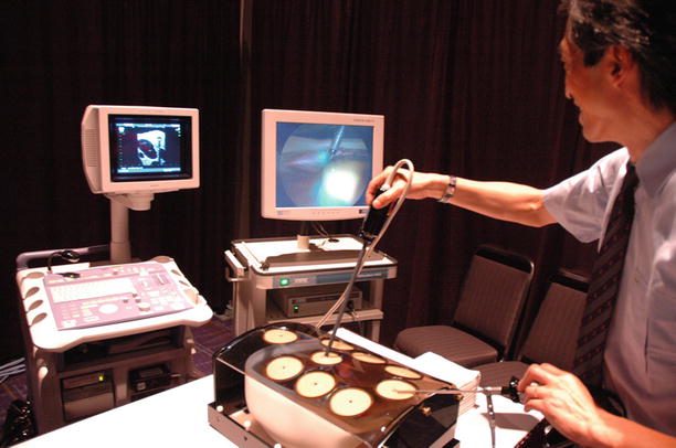
Fig. 20.3
Laparoscopic ultrasound training using intraoperative/laparoscopic ultrasound phantom, which is placed in a laparoscopic trainer box (Courtesy of Kyoto Kagaku Co., Ltd., Kyoko Japan)
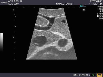
Fig. 20.4
Intraoperative ultrasound phantom scanning image: liver (Courtesy of Kyoto Kagaku Co., Ltd., Kyoko Japan)
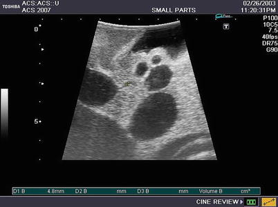
Fig. 20.5
Intraoperative ultrasound phantom scanning image: biliary system and associated surrounding structures. A cursor indicates the cystic duct (Courtesy of Kyoto Kagaku Co., Ltd., Kyoko Japan)
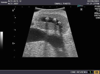
Fig. 20.6
Intraoperative ultrasound phantom scanning image: bile duct stones with shadowing (Courtesy of Kyoto Kagaku Co., Ltd., Kyoko Japan)
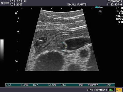
Fig. 20.7
Intraoperative ultrasound phantom scanning image: pancreas and pancreatic head tumor (cursors) (Courtesy of Kyoto Kagaku Co., Ltd., Kyoko Japan)
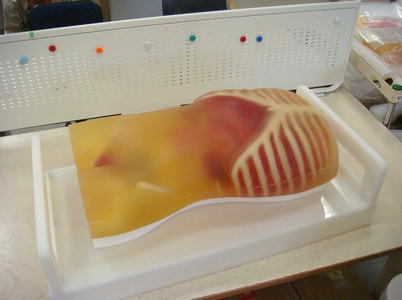
Fig. 20.8
FAST/ER phantom: abdominal/thoracic ultrasound phantom including abdominal pathology such as intra-abdominal bleeding, acute cholecystitis, and others (Courtesy of Kyoto Kagaku Co., Ltd., Kyoko Japan)
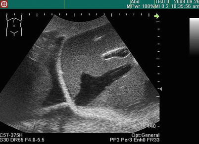
Fig. 20.9
FAST/ER phantom scanning image: cardiac tamponade (Courtesy of Kyoto Kagaku Co., Ltd., Kyoko Japan)
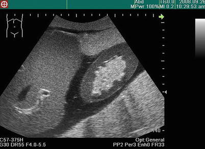
Fig. 20.10
FAST/ER phantom scanning image: intra-abdominal bleeding (Courtesy of Kyoto Kagaku Co., Ltd., Kyoko Japan)
SCAN, SCAN, SCAN!!! Ultrasound – guide, guide, guide!!! Experiences matter. How many experiences does a surgeon need for gaining ultrasound proficiency and expertise? There is no simple answer to this question. The required numbers of ultrasound scanning and interpretation experiences vary depending on the complexity of the task in each ultrasound application. Requisite experience for learning FAST is different from the learning curve for IOUS or laparoscopic ultrasound. In general, roughly 30–50 cases in which someone performs ultrasound by himself/herself may be needed to obtain confidence and competence in a specific examination. These cases should include sufficient numbers of abnormal findings. In addition to diagnostic ultrasound, ultrasound-guided procedures such as needle biopsy require probably about 15–30 cases of independent performance. Because ultrasound guidance is for invasive procedures, it is encouraged to practice such ultrasound procedures using ultrasound phantoms including target lesions before performing on patients.
Seek out opportunities to perform ultrasound. For example, if seeing a consult in the ER for possible cholecystitis, perform your own right upper quadrant ultrasound. This experience will require finding appropriate acoustic windows, identifying surrounding organs aside from the gallbladder, including the liver, kidney, and pancreas. Proper interpretation of the scans will require distinguishing between a normal and an inflamed gallbladder and gallstones versus artifact or appreciating findings suggesting alternative diagnoses. “Close the loop” by comparing your own ultrasound scans and interpretation to the formal radiology examination. If a radiologist or an expert surgeon-sonographer is available and amenable, ask for feedback.
Learn in a graduated approach – start first with simple ultrasound tasks. Mastery of basic skills is required before complex tasks can be accomplished. Learning ultrasound can be divided into five phases, paralleling the ACS Ultrasound Education Program:
Get Clinical Tree app for offline access

1.
Learning basic ultrasound concepts (cognitive)
Basic ultrasound physics (pulse-echo principle, propagation speeds, impedance, attenuation, reflection, resolution, etc.)
Basic ultrasound instrumentation (machine components)
Basic ultrasound scanning techniques (probe orientation, probe handling [rocking, rotating, sliding], ultrasound planes)
Basic ultrasound interpretation principles (machine assumptions, hypoechoic, hyperechoic, isoechoic, artifacts [shadowing, enhancement, reverberation, mirror image, etc.])
2.
Learning advanced ultrasound applications (cognitive)
Advanced concepts for specific ultrasound applicationsRelated posts:
 Introduction: The Importance of Ultrasound in a Surgical Practice
Abdominal Ultrasound: Credentialing and Certification
Introduction: The Importance of Ultrasound in a Surgical Practice
Abdominal Ultrasound: Credentialing and Certification
 Trauma Ultrasound
 Techniques in Laparoscopic and Intraoperative Ultrasound Guidance
 Abdominal Wall Anatomy, Pathology, and Intervention
 Percutaneous Ultrasound Guidance Techniques and Procedures

Stay updated, free articles. Join our Telegram channel

Full access? Get Clinical Tree

