Fig. 19.1
Granulating tissue demonstrating the healing process
The implementation in the use of BP has greatly facilitated the complex hernia treatment. This kind of ventral hernia repair represents a significant challenge for surgeons. The complexity of hernias could derive from trauma, contamination/infection, tissue loss, dimensions, anatomic position, and clinical or pharmacological data (Figs. 19.2 and 19.3). BP have completely changed the way to face the hernia surgery. They introduced the tissue engineering in surgical practice. The implantation of a biologic material triggers a cascade of events leading to new healthy tissue deposition and prosthesis remodeling. It also allows blood, growth and pro-/anti-inflammatory factors, and drugs to reach the surgical field during the first phases of healing process. This for sure enhances the effect against potential or certain contamination/infection [25]. It has been demonstrated in animal models as the tensile strength is different between cross-linked and non-cross-linked meshes during the first months after the implant. However, it reaches similar values after 12 months with the two kinds of meshes. Moreover, the strength of the repair sites doesn’t change over time. This might indicate that new tissue is deposited in the repair site as the scaffold is degraded, preventing the site from weakening over time. Another factor that should be kept into account in choosing which kind of BP to use is the demonstration that non-cross-linked material exhibits more favorable remodeling characteristics. This has a great importance when BP are used as bridge to cover tissue loss. In fact, discordant data have been published about the use of BP to bridge wide defect. Few different nonrandomized studies have been published reporting recurrence rate ranging between 100 % and 0 % if the prosthesis are placed, respectively, either as a bridge or not. Even if high-quality comparative data about BP exists in animal models, only clinical reports of a restricted number of cases are reported for humans. No definitive evidence-based conclusions could be obtained from the literature. The majority of surgeons stated they use BP in “difficult” situations, especially those with contaminated or infected field [11].
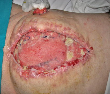
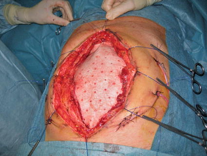
Fig. 19.2
Biological prosthesis applied in the closure of a laparotomy for trauma-related injuries; it could be utilized with vacuum-assisted closure techniques as in this case (the vacuum dispositive has been removed). To be noted is the presence of the stoma
Fig. 19.3
A biological prosthesis utilized as a bridge between fascial margins to close a laparotomy for trauma-related injuries
The Italian Biological Prosthesis Work Group (IBPWG) proposed a decisional model in the use of BP to facilitate the choice between the different types of BP [25].
The aforementioned decisional model suggests that the decision about which prosthesis to utilize should always be a dynamic process mediated by the surgeon decisional capability. The principal variables to keep in mind in deciding the kind of BP to use are infection grade and loss of tissue size.
Infection has been divided into three possible grades:
1: potentially contaminated
2: contaminated
3: infected
The same three-step division has been adopted for the tissue loss:
1: no tissue loss
2: 0–5 cm defect
3: >5 cm defect
By combining together these variables (multiplication), a score could be obtained which suggests the necessity to use either a cross-linked or a non-cross-linked BP (Figs. 19.4 and 19.5).
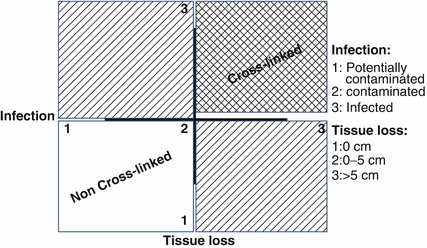
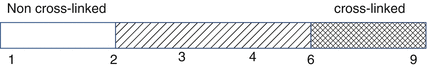
Fig. 19.4
Decisional model diagram: the product of the infection and the loss of tissue scores give as a result the value which indicates the kind of biological prosthesis to use
Fig. 19.5
Decisional line: the different results indicate the kind of biological prosthesis to use
19.2.9 Quality of Life After Abdominal Wall Reconstruction
Many studies well documented the immediate impact of injury on overall quality of life (QoL). Immediately after the injury, there is a decrease in QoL with a recovery to near baseline over a period lasting at least 1 year [26]. However, most of these studies focused more on trauma patients in general than on patients managed with open abdomens. The reconstruction of the abdominal wall after an open abdomen usually takes place during the first year after injury. At this time point, QoL and functional ability have not returned to near baseline yet. This leads to a “second hit” to QoL and functional ability. Cheatham et al. reported the long-term outcomes of open abdomen management in two papers [27]. On one hand, the authors demonstrated a decrease in QoL in both studies immediately after the first intervention, and on the other hand, they showed that patients recovered to near-normal QoL after abdominal wall reconstructive procedure. The main criticisms of the aforementioned studies are the shortness of the follow-up and the absence of measurement of depression and/or post-traumatic stress disorder. The real effect of the “second hit” on recovery might be investigated with multicenter prospective study in those centers that habitually treat these kinds of disease.
19.3 Conclusions
The best evidence available suggests that the great majority of large abdominal wall defect repair should be performed with the use of prosthetic materials as reinforcement. Due to its complexity, the abdominal wall reconstruction needs to be performed in specialized centers with a multidisciplinary approach. In contaminated or infected field, biological prosthesis should be considered as a fundamental part of the armamentarium of our surgical practice.
References
1.
Usher FC, Fries JG, Ochsner JL et al (1959) Marlex mesh, a new plastic mesh for replacing tissue defects: II. Clinical studies. Arch Surg 78:138CrossRef
Related posts:


Stay updated, free articles. Join our Telegram channel

Full access? Get Clinical Tree


